 Back
BackCell Communities: Tissues, Stem Cells, and Cancer
Study Guide - Smart Notes
 Tailored notes based on your materials, expanded with key definitions, examples, and context.
Cell Communities: Tissues, Stem Cells, and Cancer
Introduction to Cell Communities
Cells in multicellular organisms are organized into tissues, which often assemble into organs. The organization and function of tissues depend on both the cells themselves and the extracellular structures that surround them. In animals, the extracellular matrix (ECM) provides mechanical support and helps organize cells, while in plants, the cell wall fulfills a similar role.
Extracellular Matrix and Connective Tissue
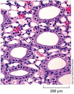
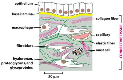
Composition and Function of Connective Tissue
Animal connective tissues are largely composed of the extracellular matrix, which varies in composition depending on the tissue type. The ECM is secreted by specialized cells such as fibroblasts, chondroblasts, and osteoblasts. Major components include:
Glycosaminoglycans (GAGs) and proteoglycans: Form hydrated gels that resist compressive forces.
Collagens: Fibrous proteins that provide tensile strength and elasticity.
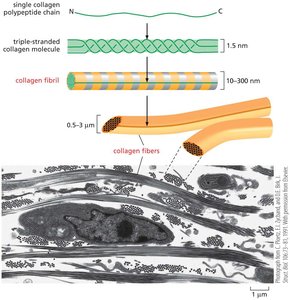
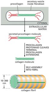
Collagen Structure and Assembly
Collagen is the most abundant protein in mammals, making up about one-quarter of total protein mass. Collagen molecules assemble into fibrils and then into larger fibers, which are organized at right angles to maximize strength. Collagen is secreted as procollagen, which has terminal peptides that prevent premature assembly. These peptides are cleaved outside the cell, allowing collagen molecules to self-assemble into fibrils.
Collagen is secreted by fibroblasts and other connective tissue cells.
Collagen fibers are aligned and organized by the cells that secrete them.

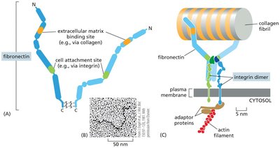
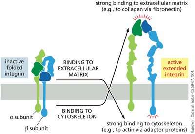
Cell-ECM Interactions: Fibronectin and Integrins
Cells interact with the ECM through fibronectin proteins, which bind collagen and connect to integrins in the plasma membrane. Integrins span the membrane and link the ECM to the cytoskeleton, allowing cells to sense and respond to their environment. Integrins can switch between inactive and active conformations, transmitting signals across the membrane.
Binding of integrins to ECM components can activate intracellular signaling pathways.
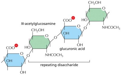
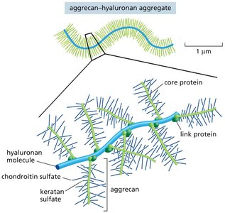
Glycosaminoglycans (GAGs) and Proteoglycans
GAGs are long, negatively charged polysaccharide chains composed of repeating disaccharide units. When attached to core proteins, they form proteoglycans. These molecules form hydrated gels that resist compression, facilitate cell migration, and regulate the passage of molecules through tissues. Hyaluronan is a major GAG that can form large aggregates with proteoglycans such as aggrecan.
Epithelial Sheets and Cell Junctions
Organization and Polarity of Epithelial Sheets
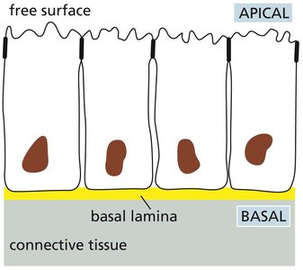
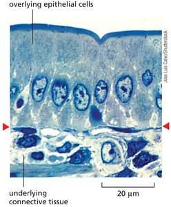
Epithelial cells form continuous sheets that cover body surfaces and line internal cavities. These sheets are polarized, with an apical surface exposed to the environment or lumen and a basal surface attached to the basal lamina (a specialized ECM layer). Cell polarity is essential for directional transport and tissue function.
Types of Cell Junctions in Epithelia
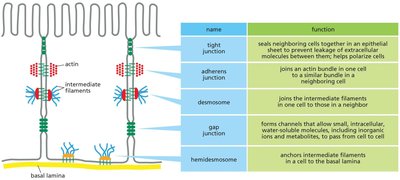
Cell junctions are specialized structures that connect epithelial cells to each other and to the basal lamina. They are essential for maintaining tissue integrity, communication, and polarity. The main types of cell junctions include:
Name | Function |
|---|---|
Tight junction | Seals neighboring cells together to prevent leakage of extracellular molecules; helps polarize cells |
Adherens junction | Joins an actin bundle in one cell to a similar bundle in a neighboring cell |
Desmosome | Joins the intermediate filaments in one cell to those in a neighbor |
Gap junction | Forms channels that allow small, intracellular, water-soluble molecules, including ions and metabolites, to pass from cell to cell |
Hemidesmosome | Anchors intermediate filaments in a cell to the basal lamina |
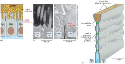
Tight Junctions
Tight junctions seal adjacent epithelial cells together, preventing the passage of molecules between them and maintaining the polarity of the epithelial sheet. They are formed by claudin and occludin proteins.
Adherens Junctions and Desmosomes
Adherens junctions and desmosomes both contain cadherin proteins, which mediate cell-cell adhesion in a calcium-dependent manner. Adherens junctions connect actin filaments, allowing coordinated contraction and tissue morphogenesis. Desmosomes connect intermediate filaments (keratin), providing mechanical strength, especially in tissues subject to stress (e.g., skin).
Hemidesmosomes
Hemidesmosomes anchor epithelial cells to the basal lamina via integrins, linking the cytoskeleton to the ECM.
Gap Junctions
Gap junctions are channels formed by connexon complexes that allow direct communication between the cytoplasms of adjacent cells. They permit the passage of ions and small molecules, enabling electrical and metabolic coupling (e.g., in cardiac muscle).
Stem Cells and Tissue Renewal
Stem Cell Properties and Niches
Stem cells are undifferentiated cells capable of self-renewal and differentiation into specialized cell types. Their fate is regulated by environmental signals and their niche—a specialized microenvironment that maintains stem cell properties through physical and chemical cues.
Stem cell populations are rare; most dividing cells are proliferating precursors.
Examples of tissues with continuous renewal: blood, skin, gut epithelium.
Examples of Tissue Renewal
Intestinal epithelium: Stem cells in the crypts continuously generate new cells that migrate and differentiate as they move up the villi.
Epidermis: Renewed from stem cells in the basal layer.
Blood: All blood cell types arise from hematopoietic stem cells in the bone marrow.
Embryonic and Induced Pluripotent Stem Cells
Embryonic stem (ES) cells are pluripotent cells derived from the early embryo that can differentiate into any cell type. Induced pluripotent stem cells (iPSCs) are generated by reprogramming differentiated somatic cells through the expression of specific transcription factors (e.g., Oct4, Sox2, Klf4, cMyc). These technologies have significant therapeutic potential for regenerative medicine and disease modeling.
Cancer: Cell Communities Gone Awry
Properties and Progression of Cancer
Cancer is characterized by uncontrolled cell proliferation and the ability to invade and colonize other tissues (metastasis). Cancer cells often display genetic instability, including abnormal chromosomes. Tumor progression involves repeated rounds of mutation, proliferation, and selection, resulting in genetically diverse cell populations within a tumor.
Benign tumors do not invade or metastasize.
Malignant tumors invade surrounding tissues and can spread to distant sites.
Cancer-Critical Genes: Oncogenes and Tumor Suppressors
Two main classes of genes are commonly mutated in cancer:
Oncogenes: Gain-of-function mutations in proto-oncogenes (normal genes that promote cell growth) can drive cancer, often in a dominant manner (one mutated copy is sufficient).
Tumor suppressor genes: Loss-of-function mutations in these genes (which normally restrain cell growth) promote cancer, usually in a recessive manner (both copies must be lost).
Pathways and Examples
Mutations in key signaling pathways (e.g., Wnt pathway, APC gene) can lead to uncontrolled proliferation. For example, inactivation of both copies of the APC tumor suppressor gene is an early event in colorectal cancer, leading to over-proliferation of gut precursor cells.
Early Detection and Treatment
Cancers are often not diagnosed until they have grown to contain hundreds of millions of cells. Early detection is critical for successful treatment. Advances in stem cell technology and understanding of cancer genetics are improving therapeutic options, including targeted therapies and regenerative medicine approaches.
Summary Table: Types of Cell Junctions
Name | Function |
|---|---|
Tight junction | Seals neighboring cells together in an epithelial sheet to prevent leakage of extracellular molecules; helps polarize cells |
Adherens junction | Joins an actin bundle in one cell to a similar bundle in a neighboring cell |
Desmosome | Joins the intermediate filaments in one cell to those in a neighbor |
Gap junction | Forms channels that allow small, intracellular, water-soluble molecules, including ions and metabolites, to pass from cell to cell |
Hemidesmosome | Anchors intermediate filaments in a cell to the basal lamina |
Additional info: For further study, review the molecular mechanisms of cell signaling (Ch.16) and cell cycle regulation (Ch.18) for more examples of proto-oncogenes and tumor suppressor genes.
