 Back
BackCell Junctions and the Extracellular Matrix: Structure, Function, and Molecular Components
Study Guide - Smart Notes
 Tailored notes based on your materials, expanded with key definitions, examples, and context.
Cell Junctions and the Extracellular Matrix
Overview of Tissue Organization
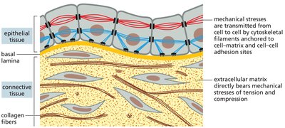
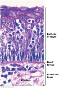
Multicellular organisms are composed of various tissues, each with specialized structural and functional properties. The organization of cells within tissues relies on cell junctions and the extracellular matrix (ECM), which together provide mechanical support, facilitate communication, and regulate cellular behavior.
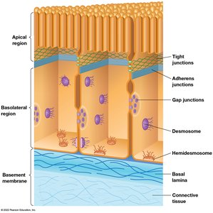
Epithelium: Sheets of polarized cells with distinct apical and basal domains.
Connective tissue: Loosely organized cells embedded in a rigid scaffold or ECM.
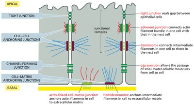
Types of Cell Junctions
Cell junctions are specialized structures that connect neighboring cells or cells to the ECM. They are essential for tissue integrity, communication, and selective permeability.
Adhesive Junctions: Mediate cell-cell adhesion; include adherens junctions and desmosomes.
Tight Junctions: Seal spaces between cells, forming a permeability barrier.
Gap Junctions: Allow exchange of ions and small molecules between cells.
Cell-Matrix Junctions: Anchor cells to the ECM (e.g., hemidesmosomes).
Adhesive Junctions
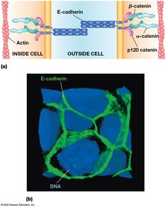
Adherens Junctions
Adherens junctions are cadherin-mediated cell-cell junctions that connect actin microfilaments in adjacent cells. They are crucial for maintaining tissue structure and facilitating cell signaling.
Cadherins: Transmembrane proteins with extracellular repeats; mediate Ca2+-dependent adhesion.
Catenins: Intracellular proteins linking cadherins to actin filaments (α-catenin, β-catenin, p120 catenin).
Function: Form continuous bands around cells, linking actin networks and supporting tissue formation.
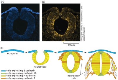
Tissue Specificity of Cadherins
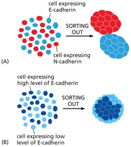
Different cadherin types are expressed in specific tissues, contributing to cell sorting and tissue specificity. E-cadherin is typical of epithelial cells, while N-cadherin is found in neurons and cardiac muscle.
Cell Sorting: Cells expressing different cadherins or levels of cadherin segregate into distinct tissues.
Development: Cadherin expression patterns guide tissue formation during embryogenesis.
Desmosomes
Desmosomes are button-like junctions providing strong adhesion between cells, especially in tissues subject to mechanical stress (e.g., skin, heart muscle). They connect intermediate filaments in adjacent cells.
Desmocollin and Desmoglein: Cadherin family proteins forming the desmosomal connection.
Adaptor Proteins: Plakoglobin, desmoplakin, plakophilin link desmosomal cadherins to intermediate filaments.
Function: Provide structural integrity and resist mechanical stress.
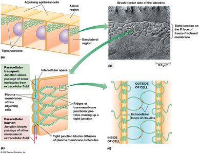
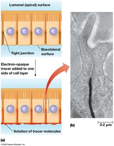
Tight Junctions
Structure and Function
Tight junctions seal the space between epithelial cells, forming a barrier to prevent movement of molecules across cell layers. They are composed of claudin proteins, which interlock to create ion-selective pores.
Claudins: Transmembrane proteins with four domains and a large extracellular loop.
Barrier Function: Restrict paracellular transport and maintain tissue compartmentalization.
No Cytoskeletal Connection: Unlike adhesive junctions, tight junctions do not connect to the cytoskeleton.
Gap Junctions
Structure and Function
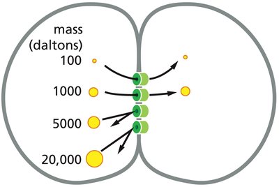
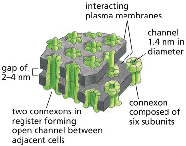
Gap junctions are channels that allow direct communication between the cytoplasm of adjacent cells. They permit the passage of ions and small molecules, enabling electrical and chemical signaling.
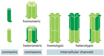
Connexins: Transmembrane proteins forming hexameric connexons.
Connexons: Two connexons from adjacent cells align to form a channel (1.4 nm diameter).
Selective Permeability: Only small molecules (< 1000 Da) can pass; larger proteins and nucleic acids are excluded.
The Extracellular Matrix (ECM)
Types of ECM
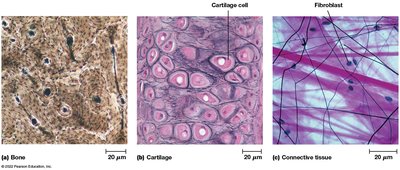
The ECM is a complex network of proteins and polysaccharides that provides structural support and regulates cellular functions. Its composition varies by tissue type.
Bone: Rigid matrix with few cells.
Cartilage: Flexible matrix with abundant proteoglycans.
Connective Tissue: Gelatinous matrix with fibroblasts and collagen fibers.
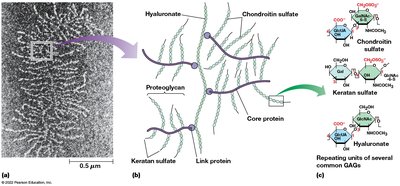
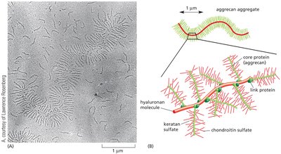
Proteoglycans and Glycosaminoglycans (GAGs)
Proteoglycans are composed of a core protein and long polysaccharide chains (GAGs). GAGs are hydrophilic, attracting water and cations to form a gelatinous matrix.
GAGs: Repeating disaccharides with amino sugars and sugar acids (e.g., chondroitin sulfate, keratan sulfate, hyaluronate).
Function: Provide hydration, support, and facilitate cell migration.
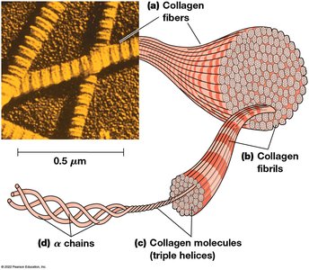
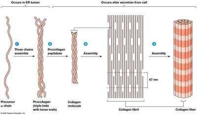
Collagen
Collagen is the most abundant ECM protein, providing tensile strength. Collagen fibers are composed of triple-helical molecules assembled into fibrils and fibers.
Structure: Three α chains form a triple helix; fibrils assemble into fibers.
Function: Resist stretching and provide structural support.
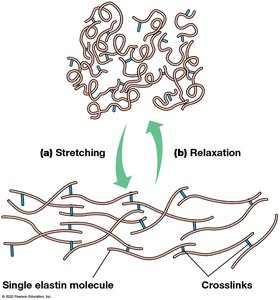
Elastin
Elastin imparts elasticity and flexibility to the ECM. Elastin molecules are crosslinked, allowing tissues to stretch and return to their original shape.
Structure: Rich in glycine and proline; crosslinked by lysine residues.
Function: Provides resilience to tissues such as skin, lungs, and blood vessels.
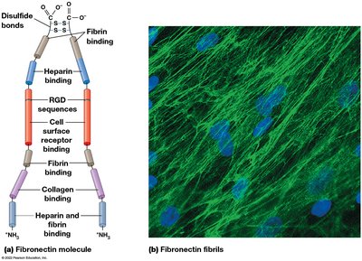
Adhesive Glycoproteins: Fibronectin and Laminin
Adhesive glycoproteins anchor cells to the ECM and facilitate cell migration, differentiation, and signaling. Fibronectin and laminin are the principal adhesive glycoproteins.
Fibronectin: Binds collagen, cell surface receptors (integrins), and fibrin; essential for cell migration and blood clotting.
Laminin: Major component of basal lamina; binds integrins and ECM molecules, supporting epithelial cell attachment.
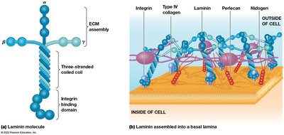


Integrins: Cell Surface Receptors
Integrin Function
Integrins are transmembrane receptors that bind ECM proteins (fibronectin, laminin) and connect the ECM to the cytoskeleton. They play a central role in cell adhesion, migration, and signaling.
Structure: Heterodimeric proteins with extracellular ligand-binding domains and intracellular cytoskeletal connections.
Function: Integrate mechanical and chemical signals from the ECM to the cell interior.
