 Back
BackCell Junctions and the Extracellular Matrix: Structure, Function, and Signaling
Study Guide - Smart Notes
 Tailored notes based on your materials, expanded with key definitions, examples, and context.
Cell Junctions and the Extracellular Matrix (ECM)
Overview of Cell Junctions
Cell junctions are specialized structures that connect cells to one another and to the extracellular matrix, playing critical roles in tissue integrity, communication, and signaling. The main types include adherens junctions, desmosomes, tight junctions, and gap junctions.
Adherens Junctions: Connect actin filaments of adjacent cells via cadherins.
Desmosomes: Link intermediate filaments using specialized cadherins.
Tight Junctions: Seal cells together to prevent passage of molecules.
Gap Junctions: Allow direct communication between cells through channels.
Cell Membrane Adhesion Proteins
Adhesion proteins mediate cell-cell and cell-matrix interactions, essential for tissue structure and signaling.
Cadherins: Mediate cell-cell adhesion; require Ca2+ for function.
Integrins: Mediate cell-matrix adhesion; involved in signaling and anchorage dependence.
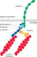
Cadherins: Structure and Function
Cadherins are a diverse family of Ca2+-dependent adhesion molecules that form homophilic bonds, contributing to cell sorting and tissue organization.
Ca2+ Binding: Stabilizes cadherin structure, preventing flexing.
Low Affinity Binding: Multiple cadherins interact, creating strong adhesion via the "Velcro" principle.
Homophilic Binding: Cadherins bind to identical cadherins on adjacent cells.
Catenins Link Cadherins to Actin Filaments
Catenins are adaptor proteins that connect cadherins to the actin cytoskeleton, enabling mechanotransduction and structural integrity.
p120-catenin, β-catenin, α-catenin: Mediate linkage between cadherins and actin filaments.
Vinculin: Reinforces the connection under mechanical stress.
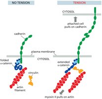
Mechanotransduction at Cell Junctions
Mechanotransduction is the process by which junction proteins sense mechanical stress and generate biochemical signals, balancing forces across the junction.
No Tension: α-catenin is folded; vinculin is not bound.
Tension: α-catenin extends, exposing binding sites for vinculin, which strengthens the junction.
Actin-Myosin Interaction: Myosin II pulls on actin, increasing tension.
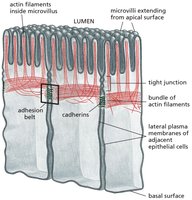
Adherens Junctions in Epithelial Cells
Adherens junctions are prominent in epithelial tissues, forming adhesion belts that maintain tissue integrity and facilitate morphogenesis.
Adhesion Belt: Encircles cells, connecting actin filaments via cadherins.
Microvilli: Supported by actin filaments, increasing surface area.
Tight Junctions: Located apically, prevent paracellular transport.
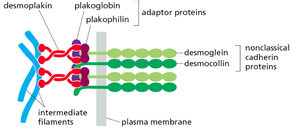
Desmosomes: Structure and Function
Desmosomes are cell-cell junctions similar to adherens junctions but connect intermediate filaments via specialized cadherins (desmoglein, desmocollin).
Intermediate Filaments: Provide mechanical strength.
Adaptor Proteins: Plakoglobin, plakophilin, and desmoplakin link cadherins to filaments.
Nonclassical Cadherins: Desmoglein and desmocollin mediate adhesion.
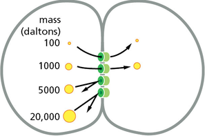
Gap Junctions: Intercellular Communication
Gap junctions are channels that allow direct exchange of ions and small molecules between adjacent cells, facilitating rapid communication.
Connexons: Protein complexes forming the channel.
Size Selectivity: Only molecules below a certain mass (e.g., 20,000 Daltons) can pass.
Function: Synchronize cellular activities, such as electrical signaling in cardiac muscle.
Extracellular Matrix (ECM)
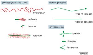
Composition of the ECM
The ECM is a complex network of proteins and polysaccharides that provides structural support, regulates cell behavior, and mediates signaling.
Proteoglycans and GAGs: Hyaluronan, perlecan, decorin, aggrecan; provide hydration and resistance to compression.
Fibrous Proteins: Collagen (type IV, fibrillar); provide tensile strength.
Glycoproteins: Laminin, nidogen, fibronectin; mediate cell adhesion and signaling.
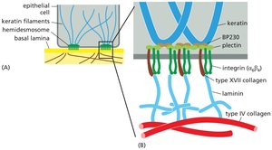
Basal Lamina (Basement Membrane)
The basal lamina is a specialized form of ECM that underlies epithelial cells and surrounds muscle, fat, and Schwann cells. It acts as a selective barrier and scaffold for tissue repair.
Composition: Laminin, type IV collagen, perlecan, nidogen.
Function: Separates cells from connective tissue, filters molecules, supports cell migration and wound repair.
Cell Contribution: Both epithelial and stromal cells secrete components.

Integrins and Cell-Matrix Adhesion
Integrins are transmembrane receptors that mediate cell-ECM adhesion and signal transduction, essential for cell survival and response to mechanical forces.
Hemidesmosomes: Specialized junctions linking keratin filaments to the basal lamina via integrins.
Anchorage Dependence: Cells undergo apoptosis if detached from the ECM.
Integrin Signaling: Can be activated from outside-in or inside-out, clustering to form adhesions.
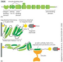
Tension Sensing and Mechanotransduction in ECM
Cells sense mechanical tension through integrins and associated proteins, triggering biochemical responses that regulate adhesion and cytoskeletal dynamics.
Talin: Unfolds under force, exposing binding sites for vinculin.
Vinculin: Binds to talin and actin, reinforcing adhesion under tension.
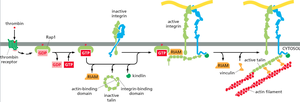
Intracellular Signaling Activates Integrins
Integrin activation involves intracellular signaling pathways that regulate their affinity for ECM ligands and linkage to the cytoskeleton.
Rap1: Small GTPase that activates integrins.
Talin and Kindlin: Bind to integrin cytoplasmic domains, promoting activation and linkage to actin filaments.
Active Integrin: Forms strong adhesions and transmits mechanical signals.
Summary Table: Cell Junctions and ECM Components
Junction Type | Main Proteins | Cytoskeletal Linkage | Function |
|---|---|---|---|
Adherens Junction | Cadherins, catenins | Actin filaments | Cell-cell adhesion, tissue integrity |
Desmosome | Desmoglein, desmocollin, plakoglobin, desmoplakin | Intermediate filaments | Mechanical strength |
Tight Junction | Claudins, occludins | Actin filaments | Barrier to paracellular transport |
Gap Junction | Connexins | None | Intercellular communication |
Hemidesmosome | Integrins, BP230, plectin | Keratin filaments | Cell-ECM adhesion |
Additional info: Academic context was added to clarify the structure and function of cell junctions, ECM components, and mechanotransduction processes. Table entries and protein details were inferred from standard cell biology knowledge.
