 Back
BackCell Membranes, Membrane Transport, Endomembrane System, and Cytoskeleton: Study Guide
Study Guide - Smart Notes
 Tailored notes based on your materials, expanded with key definitions, examples, and context.
Cell Membranes
Major Biological Functions of Cellular Membranes
Cellular membranes are essential for maintaining the integrity and functionality of cells. They:
Act as selective barriers, controlling the entry and exit of substances.
Facilitate communication via receptors and signaling molecules.
Provide structural support and compartmentalization within cells.
Enable cell recognition and adhesion.
Development of the Fluid Mosaic Model
The fluid mosaic model describes the structure of cell membranes as a dynamic combination of lipids and proteins. Key steps in its development:
Early observations of lipid bilayers (Gorter and Grendel, 1925).
Davson-Danielli model proposed a protein-lipid-protein sandwich (1935).
Freeze-fracture electron microscopy revealed proteins embedded within the bilayer (1960s).
Singer and Nicolson (1972) proposed the fluid mosaic model, highlighting lateral mobility of components.
Major Components of the Cell Membrane
Phospholipids: Form the basic bilayer structure.
Proteins: Integral and peripheral, responsible for transport, signaling, and structure.
Cholesterol: Modulates fluidity and stability.
Carbohydrates: Attached to lipids (glycolipids) or proteins (glycoproteins), involved in cell recognition.
Main Classes of Membrane Lipids and Their Structures
Phospholipids: Glycerol backbone, two fatty acids, phosphate group.
Glycolipids: Lipids with carbohydrate groups.
Sterols: Four-ring structure (e.g., cholesterol).
Phospholipids: Classification and Structure
Phospholipids include phosphoglycerides and sphingolipids.
They are amphipathic, with hydrophilic heads and hydrophobic tails.
Cholesterol: Functions and Properties
Cholesterol is a sterol that modulates membrane fluidity and permeability.
It is amphipathic, with a polar hydroxyl group and a nonpolar steroid ring structure.
Factors Influencing Membrane Fluidity
Fatty acid composition (saturated vs. unsaturated).
Cholesterol content.
Temperature.
Lipid Rafts
Lipid rafts are microdomains enriched in cholesterol, sphingolipids, and certain proteins, serving as platforms for signaling and trafficking.
Glycoproteins and Glycolipids
Glycoproteins: Proteins with attached carbohydrate chains, found on the extracellular surface.
Glycolipids: Lipids with carbohydrate groups, also on the extracellular surface.
Human Blood Types: Molecular Basis
Determined by specific glycosylation patterns on red blood cell membranes.
Type O lacks A/B antigens, making it a universal donor; type AB has both, making it a universal recipient.
Detergents and Integral Membrane Proteins
Detergents solubilize integral membrane proteins by surrounding their hydrophobic regions, keeping them soluble in aqueous solutions.
Cell Walls in Different Organisms
Plants: Cellulose-based cell walls.
Fungi: Chitin-based cell walls.
Bacteria: Peptidoglycan-based cell walls.
Key Techniques: SDS-PAGE and FRAP
SDS-PAGE: Separates proteins by size using a detergent (SDS) and polyacrylamide gel electrophoresis.
FRAP (Fluorescence Recovery After Photobleaching): Measures lateral mobility of membrane components by bleaching a fluorescent label and monitoring recovery.
Transport Across Membranes
Types of Membrane Transport
Simple Diffusion: Movement of small, nonpolar molecules down their concentration gradient without assistance.
Facilitated Diffusion: Movement of molecules down their gradient via specific transport proteins.
Passive Transport: No energy required; includes simple and facilitated diffusion.
Active Transport: Requires energy to move substances against their gradient.
Primary Active Transport: Direct use of ATP (e.g., Na+/K+ pump).
Secondary Active Transport: Uses energy from an ion gradient established by primary transport.
Symport: Two substances move in the same direction.
Antiport: Two substances move in opposite directions.
Carrier Proteins: Bind and transport specific molecules.
Channel Proteins: Form pores for passive movement of ions or water.
Osmosis: Diffusion of water across a semipermeable membrane.
Hypertonic vs. Hypotonic Solutions: Hypertonic solutions cause cells to shrink; hypotonic solutions cause swelling.
Membrane Transport in Erythrocytes
Glucose transport via GLUT1 (facilitated diffusion).
Anion exchange (Cl-/HCO3- antiport).
Osmosis regulates cell volume.
Endomembrane System
Components of the Endomembrane System
Endoplasmic reticulum (ER)
Golgi apparatus
Lysosomes
Endosomes
Transport vesicles
Plasma membrane
Rough ER vs. Smooth ER
Rough ER: Studded with ribosomes; synthesizes membrane and secretory proteins.
Smooth ER: Lacks ribosomes; involved in lipid synthesis, detoxification, and calcium storage.
Cotranslational Translocation
Process by which nascent polypeptides are inserted into the ER membrane during translation, mediated by signal sequences and the translocon.
Protein Synthesis: ER vs. Cytosol
Proteins destined for secretion, membranes, or organelles are synthesized on the ER.
Cytosolic proteins are synthesized on free ribosomes in the cytosol.
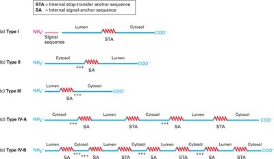
Membrane Protein Topology and Signal Sequences
Membrane protein orientation is determined by signal-anchor (SA) and stop-transfer anchor (STA) sequences, which are hydrophobic and dictate insertion and topology.
Golgi Apparatus: Structure and Function
Processes, sorts, and modifies proteins and lipids.
Cis-Golgi: Entry face, receives vesicles from ER.
Trans-Golgi: Exit face, sorts and dispatches vesicles to final destinations.
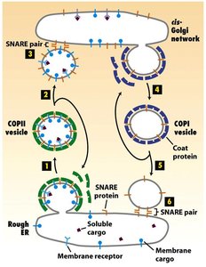
Transport Vesicles: Types and Pathways
COPII vesicles: ER to Golgi (anterograde transport).
COPI vesicles: Golgi to ER (retrograde transport).
Clathrin-coated vesicles: Golgi to endosomes, plasma membrane to endosomes.
Anterograde vs. Retrograde Transport
Anterograde: Forward movement (ER → Golgi → plasma membrane).
Retrograde: Reverse movement (plasma membrane/Golgi → ER).
Membrane Orientation During Budding and Fusion
Membrane orientation (cytosolic vs. non-cytosolic faces) is conserved during vesicle budding and fusion, ensuring proper protein and lipid topology.

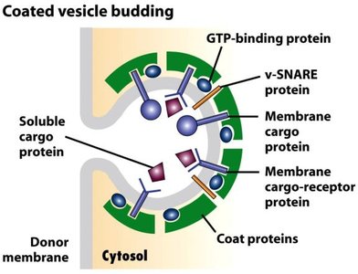
Vesicle Budding: Key Components
GTP-binding proteins: Regulate vesicle formation and targeting.
v-SNARE proteins: Mediate vesicle fusion with target membranes.
Coat proteins: Shape vesicles and select cargo.
Membrane cargo proteins: Transported proteins within vesicles.
Membrane cargo protein receptors: Recognize and bind cargo proteins.
Vesicle Coat Shedding and SNARE Exposure
Vesicle coats are shed after budding to expose v-SNAREs, which are necessary for vesicle targeting and fusion with the correct organelle membrane.
Vesicle Target Recognition
Vesicles recognize target organelles via specific interactions between v-SNAREs (vesicle) and t-SNAREs (target membrane), ensuring specificity in delivery.
Importance of Retrograde Vesicular Transport
Retrograde transport returns escaped ER proteins and recycles membrane components, maintaining organelle function and composition.
KDEL Signal and Protein Retrieval
KDEL signal: A C-terminal sequence (Lys-Asp-Glu-Leu) that directs ER-resident proteins back from the Golgi to the ER.
If a KDEL-containing protein is found in the Golgi, it is retrieved by KDEL receptors and returned to the ER via COPI vesicles.
Receptor-Mediated Endocytosis
Specific uptake of extracellular molecules via receptor binding, followed by vesicle formation and internalization.
Lysosome Functions
Degradation of macromolecules.
Recycling of cellular components.
Defense against pathogens.
Cytoskeleton
Overview of the Cytoskeleton
The cytoskeleton is a dynamic network of protein filaments that provides structural support, facilitates movement, and organizes cellular components.
Microfilaments: Components and Polarity
Composed of actin monomers (G-actin) polymerized into F-actin filaments.
Polar structures with distinct plus (+) and minus (−) ends, essential for directional movement.
Microfilaments vs. Microtubules
Microfilaments: Actin-based, ~7 nm diameter, involved in cell shape and movement.
Microtubules: Tubulin-based, ~25 nm diameter, involved in organelle movement and cell division.
Myosin: Types and Functions
Myosin is a motor protein that moves along actin filaments using ATP.
Myosin I, II, and V move toward the plus end; myosin II forms bipolar filaments for muscle contraction.
Muscle Cell Organization and Contraction
Muscle cells contain repeating units called sarcomeres, composed of thin (actin) and thick (myosin) filaments.
Contraction occurs as myosin heads pull actin filaments toward the center, shortening the sarcomere.
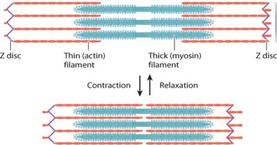
Role of Ca2+ in Muscle Contraction
Calcium ions bind to troponin, causing conformational changes that expose myosin-binding sites on actin, enabling contraction.
Microtubules: Structure and Organization
Composed of α- and β-tubulin heterodimers arranged into protofilaments, which assemble into a hollow tube.
Microtubules are polar, with a fast-growing plus end and a slow-growing minus end.
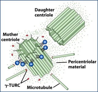
Microtubule-Organizing Centers (MTOCs) and Centrosomes
MTOCs nucleate and anchor microtubules; the centrosome is the primary MTOC in animal cells.
Non-mitotic cells have one centrosome; mitotic cells have two.
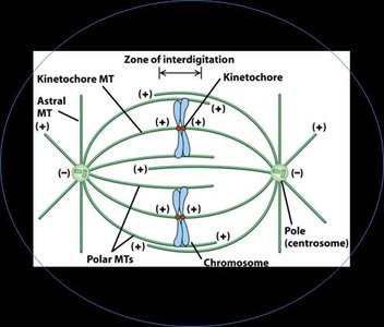
Mitotic Spindle: Microtubule Classes
Kinetochore microtubules: Attach to chromosomes.
Polar microtubules: Interact with those from the opposite pole.
Astral microtubules: Anchor spindle to cell cortex.
Dynamic Instability and Treadmilling
Dynamic instability: Microtubules rapidly switch between growth and shrinkage.
Treadmilling: Subunits add at one end and dissociate at the other, maintaining length but moving subunits through the filament.
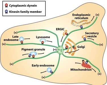
Microtubule Motor Proteins
Kinesins: Move toward the plus end (anterograde transport).
Dyneins: Move toward the minus end (retrograde transport).
Both use ATP hydrolysis for movement.
γ-Tubulin Ring Complex (γ-TuRC)
γ-TuRC nucleates microtubule assembly at the minus end, anchoring microtubules at the centrosome.
Intermediate Filaments
10 nm diameter, composed of various proteins (e.g., keratins, lamins).
Non-polar, provide mechanical strength.
No associated motor proteins.
HIV Intracellular Trafficking
HIV utilizes the cytoskeleton for intracellular movement, hijacking motor proteins for transport to and from the nucleus.
Application of Cytoskeletal Concepts
Understanding cytoskeletal dynamics is essential for interpreting cell movement, division, and intracellular transport, as well as for solving related biological problems.
