 Back
BackCell Membranes: Structure, Composition, and Function
Study Guide - Smart Notes
 Tailored notes based on your materials, expanded with key definitions, examples, and context.
Cell Membranes
Basic Organization of Membranes in Cells
The cell membrane is a fundamental structure that separates the interior of the cell from its external environment. It is present in all cells, but its complexity varies between prokaryotes and eukaryotes.
Prokaryotic cells (e.g., bacteria) possess a single plasma membrane that encloses the cell.
Eukaryotic cells have both a plasma membrane and internal membranes that compartmentalize the cell into distinct organelles.
Internal membranes in eukaryotes allow for segregation of metabolic processes.

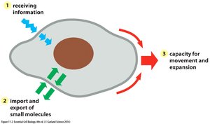
Functions of the Plasma Membrane
The plasma membrane is essential for cell communication, molecular transport, and cell growth and motility.
Receptor proteins enable the cell to receive signals from the environment.
Transport proteins facilitate the import and export of small molecules.
The membrane's flexibility and capacity for expansion allow the cell to grow, change shape, and move.
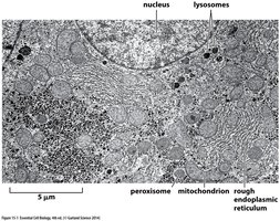
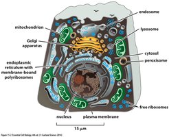
Internal Membranes and Compartmentalization in Eukaryotic Cells
Internal membranes in eukaryotic cells create enclosed compartments, segregating different metabolic processes and increasing cellular efficiency.
Major membrane-enclosed organelles include the nucleus, endoplasmic reticulum (ER), Golgi apparatus, lysosomes, endosomes, mitochondria, and peroxisomes.
Each organelle is separated from the cytosol by at least one selectively permeable membrane.
Ribosomes may be bound to the ER (rough ER) or free in the cytosol.
Structure and Composition of Cell Membranes
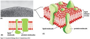
Cell Membrane Structure
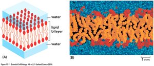
The cell membrane can be visualized in multiple ways, including electron micrographs and schematic diagrams. It consists of a lipid bilayer interspersed with protein molecules.
The lipid bilayer is approximately 5 nm thick.
Proteins are embedded within or associated with the lipid bilayer, contributing to membrane function.
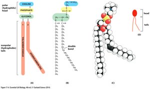
Phospholipids: The Main Component of Cell Membranes
Phosphatidylcholine is the most common phospholipid in cell membranes. Phospholipids are amphipathic molecules, containing both hydrophilic (water-loving) and hydrophobic (water-fearing) regions.
The hydrophilic head consists of choline linked to a phosphate group.
The hydrophobic tails are hydrocarbon chains (fatty acids) attached to glycerol.
A kink in one tail is caused by a double bond, affecting membrane fluidity.
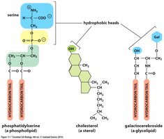
Types of Membrane Lipids
Cell membranes contain various types of amphipathic lipids:
Phospholipids (e.g., phosphatidylserine)
Sterols (e.g., cholesterol)
Glycolipids (e.g., galactocerebroside)
Each type has a hydrophilic head and one or two hydrophobic tails.
Phospholipid Bilayer Formation
Amphipathic phospholipids spontaneously form a bilayer in water, with hydrophilic heads facing outward and hydrophobic tails facing inward.
This arrangement creates a stable barrier between the cell and its environment.
The bilayer is the foundation of all biological membranes.
Bilayer Closure and Compartment Formation
Phospholipid bilayers spontaneously close to form sealed compartments, minimizing exposure of hydrophobic edges to water and creating energetically favorable structures.
This property is essential for the formation of cellular organelles and vesicles.

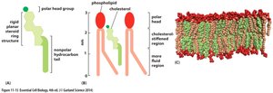
Role of Cholesterol in Membranes
Cholesterol is a sterol that intercalates between phospholipids in the bilayer, modulating membrane fluidity and stability.
Cholesterol stiffens the membrane by filling gaps between phospholipids.
It is distributed almost equally in both monolayers of the bilayer.
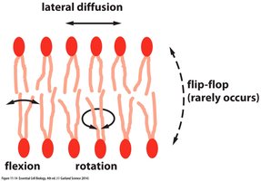
Liposomes and Membrane Motility
Pure phospholipids can form closed, spherical liposomes, which are used in research and medicine. Membrane phospholipids are motile, capable of lateral diffusion, rotation, and flexion.
Liposomes demonstrate the bilayer structure and compartmentalization.
Phospholipids rarely flip-flop between layers, maintaining membrane asymmetry.
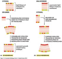
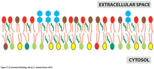
Membrane Asymmetry and Lipid Distribution
Phospholipids and glycolipids are distributed asymmetrically in the plasma membrane of eukaryotic cells.
Phosphatidylcholine and sphingomyelin are concentrated in the noncytosolic monolayer.
Phosphatidylserine and phosphatidylethanolamine are found mainly on the cytosolic side.
Glycolipids are exclusively in the noncytosolic monolayer.
Cholesterol is distributed almost equally in both monolayers.
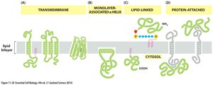
Membrane Protein Association
Membrane proteins associate with the lipid bilayer in various ways:
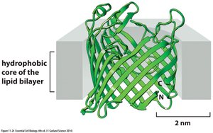
Transmembrane proteins span the bilayer as α helices or β barrels.
Monolayer-associated proteins are anchored by amphipathic α helices.
Lipid-linked proteins are attached via covalently bound lipid molecules.
Protein-attached proteins are associated through noncovalent interactions.
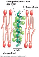
Transmembrane Protein Structure
The backbone of a polypeptide chain is hydrophilic, allowing hydrogen bonding within α helices that span the lipid bilayer.
Hydrophobic amino acid side chains interact with the lipid tails.
An α helix of about 20 amino acids is required to traverse the membrane.

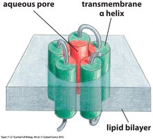
Transmembrane Pores and Channels
Transmembrane hydrophilic pores can be formed by multiple amphipathic α helices, creating water-filled channels across the bilayer.
Porin proteins form water-filled channels in bacterial outer membranes using β sheets.
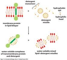
Solubilization of Membrane Proteins
Membrane proteins can be solubilized by mild detergents, which disrupt the lipid bilayer and bring proteins into solution as protein-detergent complexes.
Detergents form micelles that solubilize both proteins and lipids.
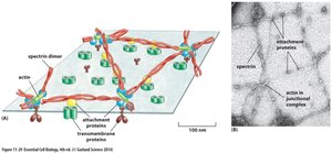
Cell Cortex and Membrane Shape
The cell cortex, composed of a spectrin meshwork, supports the plasma membrane and maintains cell shape, especially in red blood cells.
Spectrin dimers form tetramers, which, together with actin, create a mesh attached to the membrane.
This structure is essential for the characteristic biconcave shape of red blood cells.
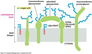
Glycocalyx: Sugar Coating of Eukaryotic Cells
Eukaryotic cells are coated with sugars, forming the glycocalyx. This carbohydrate layer is made of oligosaccharide side chains attached to membrane glycolipids and glycoproteins, and polysaccharide chains on proteoglycans.
All carbohydrate is located on the external (noncytosolic) surface of the plasma membrane.
The glycocalyx is important for cell recognition, protection, and adhesion.
Restriction of Membrane Protein Mobility
The lateral mobility of plasma membrane proteins can be restricted by various mechanisms:
Proteins can be tethered to the cell cortex, extracellular matrix, or proteins on another cell.
Diffusion barriers can restrict proteins to specific membrane domains.
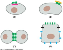
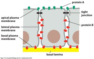
Membrane Domains in Epithelial Cells
Membrane proteins are restricted to particular domains of the plasma membrane in epithelial cells, such as those in the gut.
Protein A is found in the apical membrane, while protein B is in the basal and lateral membranes.
Tight junctions prevent proteins from diffusing between domains.
The basal lamina supports epithelial sheets.
Summary Table: Examples of Plasma Membrane Proteins and Their Functions
Functional Class | Protein Example | Specific Function |
|---|---|---|
Transporters | Na+ pump | Actively pumps Na+ out of cells and K+ in |
Ion channels | K+ leak channel | Allows K+ ions to leave cells, affecting cell excitability |
Anchors | Integrins | Link intracellular actin filaments to extracellular matrix proteins |
Receptors | PDGF receptor | Binds extracellular PDGF, generating intracellular signals |
Enzymes | Adenylyl cyclase | Catalyzes production of cAMP in response to signals |
Summary Table: Main Functions of Membrane-Enclosed Compartments in Eukaryotic Cells
Compartment | Main Function |
|---|---|
Cytosol | Contains many metabolic pathways, protein synthesis, cytoskeleton |
Nucleus | Contains main genome, DNA and RNA synthesis |
Endoplasmic reticulum (ER) | Synthesis of most lipids, proteins for distribution to organelles and plasma membrane |
Golgi apparatus | Modification, sorting, and packaging of proteins and lipids |
Lysosomes | Intracellular degradation |
Endosomes | Sorting of endocytosed material |
Mitochondria | ATP synthesis by oxidative phosphorylation |
Chloroplasts | ATP synthesis and carbon fixation by photosynthesis |
Peroxisomes | Oxidation of toxic molecules |
Summary Table: Relative Volumes and Numbers of Major Membrane-Enclosed Organelles in a Liver Cell (Hepatocyte)
Intracellular Compartment | Percentage of Total Cell Volume | Approximate Number per Cell |
|---|---|---|
Cytosol | 54 | 1 |
Mitochondria | 22 | 1700 |
Endoplasmic reticulum | 12 | 1 |
Nucleus | 6 | 1 |
Golgi apparatus | 3 | 1 |
Peroxisomes | 1 | 400 |
Lysosomes | 1 | 300 |
Endosomes | 1 | 200 |
Key Equations and Concepts
Phospholipid Bilayer Formation
The spontaneous formation of a bilayer is driven by the amphipathic nature of phospholipids:
Membrane Protein Structure
Transmembrane α helices are stabilized by hydrogen bonds:
Membrane Fluidity
Cholesterol modulates membrane fluidity:
Summary
Cell membranes are complex structures composed of lipids, proteins, and carbohydrates. Their organization and composition are essential for cellular compartmentalization, communication, transport, and structural integrity.
