 Back
Cell Signaling and Signal Transduction: Structured Study Notes for Cell Biology
Study Guide - Smart Notes
 Tailored notes based on your materials, expanded with key definitions, examples, and context.
Cell Signaling: Overview
Introduction to Cell Signaling
Cell signaling is a fundamental process by which cells communicate with each other and respond to their environment. This communication is essential for regulating cellular activities, coordinating multicellular functions, and maintaining homeostasis. - Key Point 1: Signals can originate from within the cell, from other cells, or from the external environment. - Key Point 2: The main goal of signaling is to alter the actions of target cells via chemical messengers.
Types of Cell Signaling
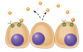
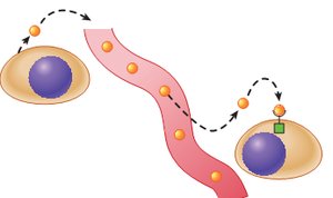
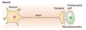
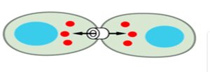
Cells utilize several distinct signaling mechanisms to communicate: - Autocrine signaling: Cells release signals that act on themselves. - Paracrine signaling: Signals affect nearby cells. - Endocrine signaling: Hormones travel through the bloodstream to distant cells. - Cell-to-cell contact: Direct communication via gap junctions. - Electrical signaling: Transmission via synapses in neurons. 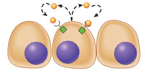
Coordination and Effects of Signaling Molecules
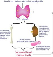
- Synergistic effect: Two ligands work together to produce a greater effect than either alone (e.g., FSH and testosterone in sperm production). - Permissive effect: One ligand enhances the responsiveness of a target organ to another hormone (e.g., PTH enhances vitamin D3 action). - Antagonistic effect: One hormone inhibits the effect of another (e.g., insulin promotes fat formation, glucagon promotes fat breakdown). 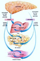
Signal Transduction Pathways
Basic Elements of Cell Signaling Cascades
Signal transduction begins with the release of a messenger molecule, which binds to a specific receptor. This binding causes a conformational change in the receptor, relaying the signal across the membrane and activating intracellular signaling proteins or enzymes. - Key Point: Second messengers are generated to amplify and propagate the signal.
Signaling Molecules
Chemical messengers transmit information between cells and are classified based on their origin and function: - Nervous system: Neurotransmitters (e.g., acetylcholine, dopamine) - Endocrine system: Hormones (e.g., insulin, glucagon) - Immune system: Cytokines (e.g., IL-6, TGFβ) - Other types: Amino acid derivatives, steroids, gases (NO), eicosanoids, polypeptides, antigens
Control of Release of Signaling Molecules
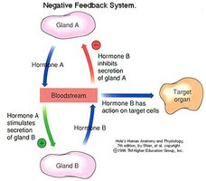
The release of extracellular signaling molecules is regulated by: - Neural control - Hormonal control - Nutrient levels (e.g., amino acids, glucose) - Ion control (e.g., Ca2+) - Antigen challenge (e.g., NF-κB activation) 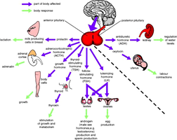
Cell Surface Receptors
Structure and Function
Cell surface receptors are proteins with a binding site for a specific chemical messenger. They act as signal transducers, with extracellular, membrane-spanning, and intracellular domains. - Types: G-Protein Coupled Receptors (GPCRs), Enzyme-linked receptors (RTKs), Ion channel-linked receptors 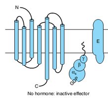
G-Protein Coupled Receptors (GPCRs)
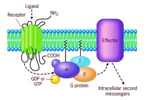
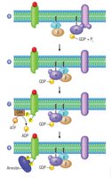
GPCRs are the largest family of cell-surface receptors, characterized by seven transmembrane domains and heterotrimeric G proteins (α, β, γ subunits). - Ligands: Hormones, neurotransmitters, chemoattractants, odorants, tastants, photons - Activation: Ligand binding causes GDP release from the α subunit, GTP binding, and dissociation of Gα from Gβγ. 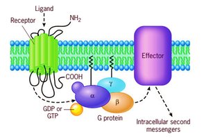
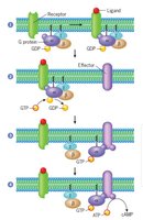
GPCR Signaling Mechanism
GPCRs activate effectors such as adenylyl cyclase and phospholipase C via G proteins: - Gαs: Activates adenylyl cyclase - Gαi: Inhibits adenylyl cyclase - Gαq: Activates phospholipase C - Gα12: Function not fully elucidated 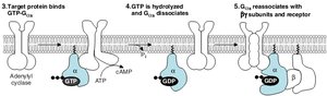
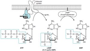
Signal Amplification: Adenylyl Cyclase and cAMP
The binding of a single hormone molecule can activate adenylyl cyclase, producing many cAMP molecules. cAMP acts as a second messenger, synthesized from ATP and degraded by phosphodiesterase. - Equation: 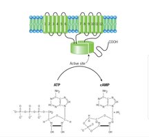
Signal Amplification: Phospholipase C Pathway
GPCRs can activate phospholipase C, which splits inositol phospholipid into: - IP3: Opens ER Ca2+ channels, triggering secretion and muscle contraction - Diacylglycerol (DAG): Activates protein kinase C (PKC), which phosphorylates target proteins 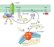
Receptor Tyrosine Kinases (RTKs)
Structure and Function
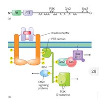
RTKs are enzymes that phosphorylate tyrosine residues on protein substrates. They have extracellular, transmembrane, and intracellular catalytic domains. Hormone binding activates kinase activity, creating docking sites for signaling proteins and initiating downstream cascades. 
Ligands and Cellular Effects
RTKs regulate growth, division, differentiation, survival, attachment, and migration. Ligands include insulin, IGF1, EGF, GH, PDGF. 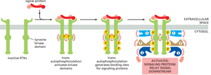
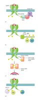
Insulin Receptor (IR)
The insulin receptor is a protein-tyrosine kinase composed of α and β chains, held together by disulfide bonds. - α chain: Extracellular, insulin-binding site - β chain: Extracellular, transmembrane, and cytoplasmic regions 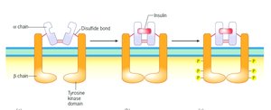
Intracellular Receptors
Structure and Function
Intracellular receptors bind lipophilic messengers (Class I hormones) such as steroid and thyroid hormones. These receptors are located in the cytosol or nucleus. Hormone-receptor complexes bind DNA, regulating gene transcription. 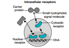
Glucocorticoid and Thyroid Hormone Signaling
- Glucocorticoids: Bind cytosolic receptors, activate gene transcription, increase blood glucose, stimulate proteolysis, inhibit muscle protein synthesis, increase fat mobilization, and modulate immune responses. - Thyroid hormones: Nuclear receptors already bound to DNA; ligand binding dissociates corepressors and activates gene transcription.
Summary Table: Types of Cell Signaling
Type | Distance | Example |
|---|---|---|
Autocrine | Self | Immune cell cytokine release |
Paracrine | Nearby | Growth factors in tissue repair |
Endocrine | Distant | Insulin, thyroid hormone |
Cell-to-cell contact | Adjacent | Gap junctions |
Electrical | Synaptic | Neurotransmitter release |
Clinical Correlates
Cholera Toxin
Cholera toxin causes ADP ribosylation of the Gα subunit, preventing GTP hydrolysis and leading to prolonged cAMP elevation, resulting in severe diarrhea.
Diabetes Mellitus
Defects in insulin signaling can lead to Type 1 or Type 2 diabetes, affecting glucose metabolism and cellular responses.
Conclusion
Cell signaling and signal transduction are essential for cellular communication, regulation, and response to environmental cues. Understanding these pathways is fundamental for cell biology and medical sciences. Additional info: Academic context was added to clarify mechanisms, clinical relevance, and molecular details for exam preparation.
