 Back
Back20: Cell Signaling Pathways: G Proteins, GPCRs, and RTKs
Study Guide - Smart Notes
 Tailored notes based on your materials, expanded with key definitions, examples, and context.
Cell Signaling Mechanisms
Intracellular Signaling Proteins as Molecular Switches
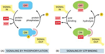
Intracellular signaling proteins often function as molecular switches, toggling between active and inactive states in response to cellular signals. Two major mechanisms are phosphorylation and GTP binding.
Phosphorylation: Protein kinases add phosphate groups (from ATP) to proteins, activating them, while protein phosphatases remove these groups, inactivating the proteins.
GTP Binding: GTP-binding proteins (G proteins) switch between active (GTP-bound) and inactive (GDP-bound) states. GTP hydrolysis inactivates the protein.
Example: Many signaling cascades in cells rely on these molecular switches to propagate and regulate signals.
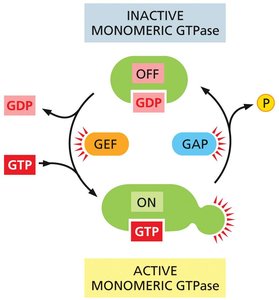
Regulation of Monomeric GTPases
Monomeric GTPases are regulated by two types of accessory proteins:
GEFs (Guanine nucleotide Exchange Factors): Promote the release of GDP, allowing GTP to bind and activate the GTPase.
GAPs (GTPase-Activating Proteins): Stimulate GTP hydrolysis, returning the GTPase to its inactive state.
Example: The Ras protein, a key regulator in cell growth, is controlled by GEFs and GAPs.
G Protein-Coupled Receptors (GPCRs)
Structure and Function of GPCRs
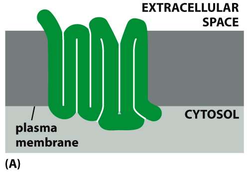
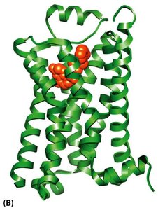
GPCRs are a large family of cell-surface receptors characterized by seven transmembrane α-helices. They detect a wide variety of signals, including hormones, neurotransmitters, and sensory stimuli.
Found in diverse organisms, including yeast and humans.
Largest family of cell-surface receptors in animals.
Example: Human olfactory and photoreceptor cells use GPCRs to detect odors and light, respectively.
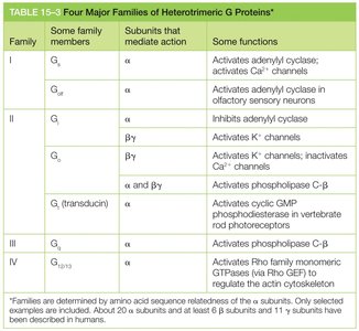
G Proteins: Structure and Activation
G proteins are heterotrimeric proteins composed of α, β, and γ subunits. The α and γ subunits are membrane-tethered. The α subunit binds GDP/GTP and has intrinsic GTPase activity.
Inactive when GDP-bound; active when GTP-bound.
Activation involves exchange of GDP for GTP, often triggered by GPCRs.
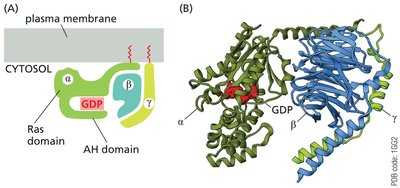
Example: The Ras domain within the α subunit is critical for GTP binding and hydrolysis.
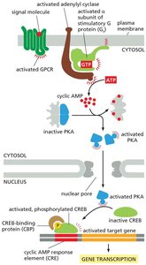
Activation of G Proteins by GPCRs
Upon ligand binding, GPCRs undergo a conformational change, activating the associated G protein by promoting GDP-GTP exchange on the α subunit. The GTP-bound α subunit and the βγ complex can then regulate downstream effectors.
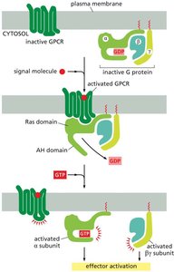
Example: Activated G proteins can open ion channels or activate enzymes such as adenylyl cyclase and phospholipase C.
Second Messenger Pathways
cAMP Synthesis and Degradation
Adenylyl cyclase catalyzes the conversion of ATP to cyclic AMP (cAMP), a key second messenger. cAMP is rapidly degraded to 5'-AMP by phosphodiesterases, ensuring transient signaling.
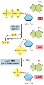
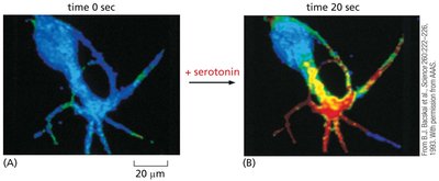
Example: The neurotransmitter serotonin increases cAMP levels in nerve cells, leading to rapid cellular responses.
Activation of Protein Kinase A (PKA) by cAMP
cAMP binds to the regulatory subunits of PKA, releasing the active catalytic subunits. These subunits can enter the nucleus and phosphorylate transcription factors such as CREB, altering gene expression.
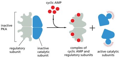
Example: cAMP-mediated activation of PKA regulates glycogen breakdown in muscle and liver cells.
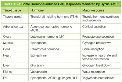
Hormone-Induced Cell Responses Mediated by cAMP
Various hormones utilize cAMP as a second messenger to elicit specific cellular responses in different tissues.
Target tissue | Hormone | Major response |
|---|---|---|
Thyroid gland | Thyroid-stimulating hormone (TSH) | Thyroid hormone synthesis and secretion |
Adrenal cortex | Adrenocorticotropic hormone (ACTH) | Cortisol secretion |
Ovary | Luteinizing hormone (LH) | Progesterone secretion |
Muscle | Epinephrine | Glycogen breakdown |
Bone | Parathyroid hormone | Bone resorption |
Heart | Epinephrine | Increase in heart rate and force of contraction |
Liver | Glucagon | Glycogen breakdown |
Kidney | Vasopressin | Water resorption |
Fat | Epinephrine, ACTH, glucagon, TSH | Triglyceride breakdown |
Phospholipase C Pathway and Calcium Signaling
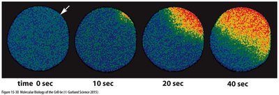
Phospholipase C-β (PLCβ) hydrolyzes PI(4,5)P2 to generate two second messengers: diacylglycerol (DAG) and inositol 1,4,5-trisphosphate (IP3). DAG remains in the membrane and activates protein kinase C, while IP3 diffuses into the cytosol and triggers Ca2+ release from the endoplasmic reticulum.
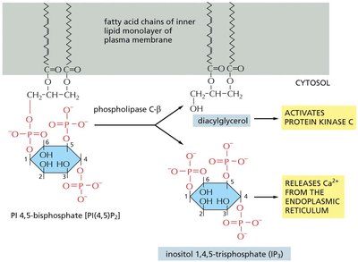
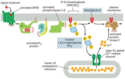
Example: The rise in cytosolic Ca2+ activates calmodulin and CaM-dependent kinases, regulating diverse cellular processes.
Calmodulin and Ca2+-Dependent Protein Kinases
Calmodulin is a ubiquitous Ca2+-binding protein that undergoes a conformational change upon binding Ca2+. It activates various target proteins, including CaM kinases and Ca2+-ATPases.
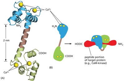
Example: CaM kinases play critical roles in learning and memory by phosphorylating synaptic proteins.
GPCR-Mediated Sensory Signaling
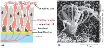
Olfactory Receptors and Smell
Olfactory receptors are GPCRs that detect odorants and activate Golf proteins, leading to increased cAMP and opening of cAMP-gated ion channels. This results in Na+ influx and depolarization of olfactory neurons, generating nerve impulses.
Example: Each olfactory neuron expresses one type of olfactory receptor, allowing discrimination of a wide range of odors.
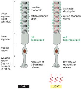
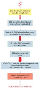
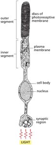
Photoreceptors and Vision
Rod photoreceptors in the retina use the GPCR rhodopsin to detect light. In darkness, cGMP levels are high, keeping cGMP-gated channels open. Light activation decreases cGMP, closing the channels and hyperpolarizing the cell.
Example: This mechanism enables vision in low-light conditions and is one of the fastest G-protein-mediated responses in vertebrates.
Enzyme-Coupled Receptors: Receptor Tyrosine Kinases (RTKs)
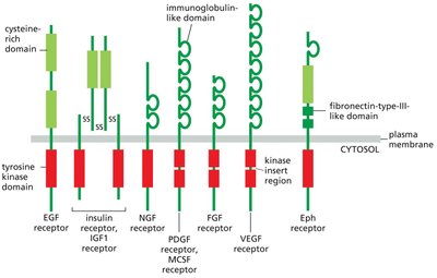
Structure and Activation of RTKs
RTKs are single-pass transmembrane proteins with an extracellular ligand-binding domain and an intracellular tyrosine kinase domain. Ligand binding induces dimerization and autophosphorylation, creating docking sites for signaling proteins.
Example: RTKs mediate responses to growth factors such as EGF, insulin, and PDGF.
RTK Signaling via Ras and MAP Kinase Pathway
Activated RTKs recruit adaptor proteins (e.g., Grb2) and Ras GEFs (e.g., Sos), leading to activation of the small GTPase Ras. Ras then initiates a kinase cascade (MAP kinase pathway) that regulates gene expression and cell division.
Example: Mutations in Ras are found in ~30% of human tumors, highlighting its importance in cell proliferation control.
MAP Kinase Modules and Scaffold Proteins
MAP kinase modules can be organized by scaffold proteins, ensuring specificity and efficiency of signaling. Different modules can mediate distinct cellular responses, such as mating or osmoregulation in yeast.
Example: Scaffold proteins prevent cross-talk between parallel MAP kinase pathways.
Summary Table: Ras Superfamily of Monomeric GTPases
Family | Some family members | Some functions |
|---|---|---|
Ras | H-Ras, K-Ras, N-Ras | Relay signals from RTKs |
Rheb | Rheb | Activates mTOR to stimulate cell growth |
Rap1 | Rap1 | Activated by a cyclic-AMP-dependent GEF; influences cell adhesion by activating integrins |
Rho* | Rho, Rac, Cdc42 | Relay signals from surface receptors to the cytoskeleton and elsewhere |
ARF* | ARF1–ARF6 | Regulate assembly of protein coats on intracellular vesicles |
Rab* | Rab1–60 | Regulate intracellular vesicle traffic |
Ran* | Ran | Regulates mitotic spindle assembly and nuclear transport of RNAs and proteins |
Additional info: The Rho, ARF, Rab, and Ran families are discussed in detail in other chapters related to cytoskeleton, vesicle transport, and nuclear transport.
