 Back
BackCell Theory, Microscopy, and Cell Types: Foundational Concepts in Cell Biology
Study Guide - Smart Notes
 Tailored notes based on your materials, expanded with key definitions, examples, and context.
Cell Theory
Historical Foundations of Cell Theory
The Cell Theory is a fundamental principle in biology, establishing the cell as the basic unit of life. Developed in the 19th century, it provides the framework for understanding biological organization and processes.
All organisms consist of one or more cells (Schwann, 1839): This principle highlights the universality of cells in living systems.
The cell is the basic unit of structure for all organisms (Schwann, 1839): Cells are the smallest entities that retain the characteristics of life.
All cells arise from preexisting cells (Virchow, 1855): This concept, "Omnis cellula e cellula," emphasizes continuity in life through cellular reproduction.
Definition of a Theory in Science
A scientific theory is a well-substantiated explanation of some aspect of the natural world, based on a body of evidence and repeatedly tested and confirmed through observation and experimentation.
Microscopy and Cell Visualization
Resolution in Microscopy
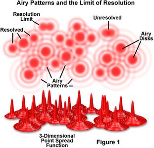
Resolution is the ability of a microscope to distinguish two points as separate entities. It is a critical parameter in cell biology, determining the level of detail observable in cellular structures.
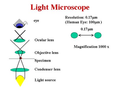
Optical (Light) Microscopes: Resolution is limited by the wavelength of light, typically about 0.2 microns ( where is the wavelength).
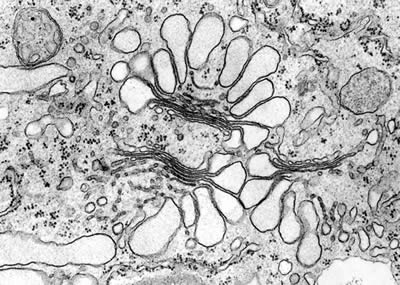
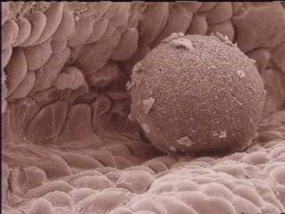
Electron Microscopes: Use electron beams, achieving much higher resolution (TEM: 0.2–0.5 nm; SEM: 10 nm).
Limitations and Improvements in Light Microscopy
Only dark objects are seen well.
Resolution is limited to about 0.2 microns.
Out-of-focus light reduces clarity.
Types of Microscopy
Different microscopy techniques are used to visualize cells and their components, each with unique advantages:
Brightfield: Uses transmitted light; best for stained specimens.
Phase Contrast: Enhances contrast in unstained cells.
Fluorescence: Visualizes specific molecules using fluorescent dyes.
Confocal: Uses lasers for high-resolution, 3D imaging.
Electron Microscopy
Transmission Electron Microscopy (TEM): Provides high-resolution images of internal cell structures.
Scanning Electron Microscopy (SEM): Visualizes cell surfaces in detail.
Cell Types and Structure
Prokaryotic vs. Eukaryotic Cells
Cells are classified into two main types based on their structural features:
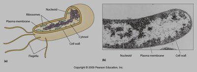
Prokaryotic Cells: Lack membrane-bound organelles and a nuclear envelope. Examples include bacteria.
Eukaryotic Cells: Possess membrane-bound organelles, including a nucleus. Examples include plant and animal cells.
Animal Cells
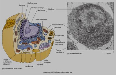
Animal cells are characterized by the absence of a cell wall, presence of lysosomes, and small vacuoles. They often have centrioles and can change shape.
Plant Cells
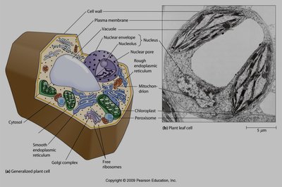
Plant cells have a fixed shape due to the cell wall, contain chloroplasts and a large central vacuole, and rarely possess cilia or flagella.
Comparison Table: Plant Cell vs. Animal Cell
Feature | Plant Cell | Animal Cell |
|---|---|---|
Shape | Fixed | Round or irregular, can change |
Cilia, flagella | Very rare | Present |
Chloroplasts and other plastids | Yes | No |
Cell wall | Yes | No |
Lysosomes | Not found | Present in cytoplasm |
Vacuoles | One large | Small in cytoplasm |
Centrioles | Only in lower plants | Yes |
Additional info:
Microscopy is essential for visualizing cells and their organelles, which are often below the resolution of the unaided eye.
Cell theory underpins all modern biological research, including genetics, molecular biology, and biotechnology.
