 Back
BackCellular Communication: Signal Transduction and Receptors
Study Guide - Smart Notes
 Tailored notes based on your materials, expanded with key definitions, examples, and context.
Cellular Communication
Introduction to Cell Signaling
Cellular communication is essential for coordinating activities in multicellular organisms. Cells use chemical signals to communicate with each other and respond to their environment. These signals are detected by specific receptors, which then trigger a cascade of intracellular events leading to a cellular response.
Chemical Signals and Cell Receptors
Types of Chemical Messengers
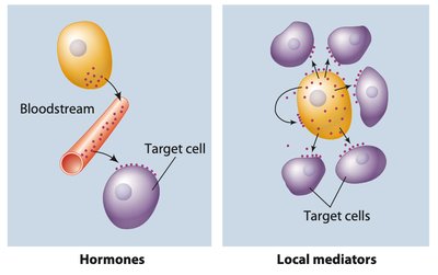
Hormones: Chemical messengers produced by specialized cells, travel through the bloodstream to distant target cells. This is known as endocrine signaling.
Local Mediators: Messengers that act near their site of production. These include:
Paracrine signals: Affect nearby cells.
Juxtacrine signals: Require direct cell-to-cell contact.
Autocrine signals: Act on the same cell that produced them.
Ligands and Receptors
Ligands: Chemical messengers that bind to receptors (e.g., hormones, neurotransmitters).
Receptors: Proteins, usually on the cell surface, that specifically bind ligands and initiate a cellular response.
Binding is determined by the specific shape and charge of the ligand and receptor, involving non-covalent interactions.
Receptor-Ligand Binding and Signal Transduction
Mechanism of Signal Transduction
When a ligand binds to its receptor, it triggers a series of intracellular events known as a signal transduction pathway. This often involves the production of secondary messengers and leads to changes in cell activity or gene expression.
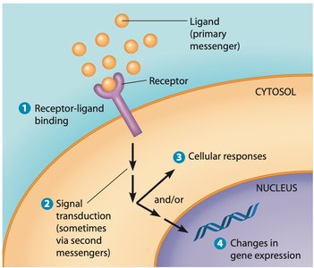
Primary messenger: The ligand itself.
Secondary messengers: Small molecules produced inside the cell in response to receptor activation (e.g., cAMP, Ca2+).
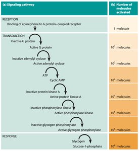
Signal amplification: A single ligand-receptor interaction can activate multiple downstream molecules, amplifying the response.
Receptor Regulation: Agonists, Antagonists, and Desensitization
Agonists and Antagonists
Agonists: Molecules that bind to receptors and activate them, mimicking the effect of the natural ligand.
Antagonists: Molecules that bind to receptors but do not activate them, blocking the action of the natural ligand.
Desensitization and Down-Regulation
Prolonged exposure to high concentrations of a ligand can lead to desensitization, where the cell reduces its response to the signal. This can involve changes in receptor number or chemistry (down-regulation).

Receptor Affinity and Dissociation Constant (Kd)
Affinity and Kd
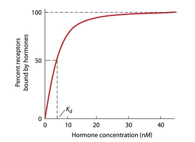
Affinity: The strength with which a receptor binds its ligand.
Dissociation constant (Kd): The concentration of ligand at which half of the receptors are occupied. Lower Kd indicates higher affinity.
Classes of Cell Surface Receptors
Major Receptor Types
Ligand-gated ion channels: Open or close in response to ligand binding, allowing ions to flow across the membrane.
G protein-coupled receptors (GPCRs): Activate G proteins, which then trigger downstream signaling pathways.
Receptor kinases: Possess intrinsic enzymatic activity, often phosphorylating tyrosine, serine, or threonine residues on target proteins.
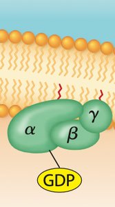
G Protein-Coupled Receptors (GPCRs) and G Proteins
Structure and Function of GPCRs
GPCRs have seven transmembrane alpha-helices.
Ligand binding activates an associated G protein, which acts as a molecular switch (on/off) depending on whether it is bound to GDP (inactive) or GTP (active).
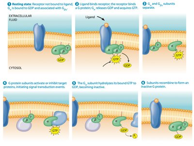
G Protein Activation Cycle
Ligand binds to GPCR, causing a conformational change.
G protein exchanges GDP for GTP on the alpha subunit.
Alpha subunit dissociates from beta and gamma subunits.
Active subunits regulate target proteins.
GTP is hydrolyzed to GDP, inactivating the G protein.
Subunits recombine to form the inactive G protein.
Second Messenger Pathways
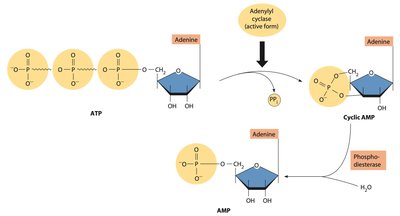
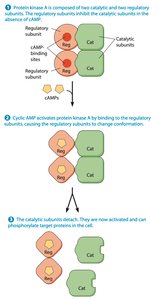
Cyclic AMP (cAMP) Pathway
cAMP is synthesized from ATP by adenylyl cyclase, which is activated by Gα-GTP.
cAMP activates protein kinase A (PKA), which phosphorylates various target proteins.
Phosphodiesterase degrades cAMP, terminating the signal.

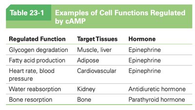
Examples of Cell Functions Regulated by cAMP
Regulated Function | Target Tissues | Hormone |
|---|---|---|
Glycogen degradation | Muscle, liver | Epinephrine |
Fatty acid production | Adipose | Epinephrine |
Heart rate, blood pressure | Cardiovascular | Epinephrine |
Water reabsorption | Kidney | Antidiuretic hormone |
Bone resorption | Bone | Parathyroid hormone |
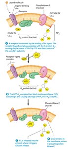
IP3/DAG Pathway
Gα-GTP activates phospholipase C, which cleaves PIP2 into IP3 and DAG.
IP3 triggers Ca2+ release from the endoplasmic reticulum.
DAG activates protein kinase C (PKC).
Calcium Ions and Calmodulin
Role of Ca2+ in Cell Signaling
Ca2+ acts as a secondary messenger in many signaling pathways.
Calmodulin is a protein that binds Ca2+ and activates various kinases and phosphatases.
Nitric Oxide (NO) Signaling
NO as a Messenger
NO is a gaseous signaling molecule that diffuses across membranes.
It activates guanylyl cyclase, increasing cGMP levels and leading to smooth muscle relaxation.
NO signaling is important in vasodilation and is targeted by drugs such as nitroglycerin and sildenafil (Viagra).
Protein Kinase-Associated Receptors
Receptor Tyrosine Kinases (RTKs)
RTKs are single-pass transmembrane proteins with intrinsic kinase activity.
Ligand binding induces dimerization and autophosphorylation of tyrosine residues.
Phosphorylated RTKs initiate signaling cascades that regulate cell growth, division, and differentiation.
Ras Pathway
Ras is a monomeric G protein activated by RTKs via guanine-nucleotide exchange factors (e.g., Sos).
Active Ras triggers a phosphorylation cascade involving Raf, MEK, and MAP kinases, ultimately regulating gene expression.
Mutations in Ras can lead to uncontrolled cell growth and cancer.
Integration of Signaling Pathways
Cell signaling pathways are highly integrated, allowing cells to process multiple signals and coordinate complex responses. Crosstalk between pathways ensures precise regulation of cellular activities.
Summary Table: Major Classes of Cell Surface Receptors
Receptor Type | Ligand Example | Mechanism | Cellular Response |
|---|---|---|---|
Ligand-gated ion channel | Acetylcholine | Ion flow across membrane | Membrane depolarization |
GPCR | Epinephrine | G protein activation, second messengers | Metabolic changes |
Receptor kinase | Growth factors | Autophosphorylation, kinase cascade | Gene expression changes |
