 Back
BackCellular Energy Metabolism, Extracellular Matrix, and Cell Junctions
Study Guide - Smart Notes
 Tailored notes based on your materials, expanded with key definitions, examples, and context.
Glycolysis and Cellular Energy Metabolism
Overview of Glycolysis
Glycolysis is a fundamental metabolic pathway in cell biology, responsible for breaking down glucose to generate energy in the form of ATP. It occurs in the cytoplasm and is the first step in chemotropic energy metabolism.
Catabolism: The breakdown of complex molecules into simpler ones, releasing energy.
Anabolism: The synthesis of complex molecules from simpler ones, requiring energy input.
ATP: The primary energy carrier in cells, with energy stored mainly in the third phosphate bond.
Phases of Glycolysis
Investment Phase: Glucose is phosphorylated twice, consuming 2 ATP molecules.
Cleavage Phase: The 6-carbon sugar is split into two 3-carbon molecules.
Energy Harvest Phase: 2 NADH and 4 ATP are produced (net gain: 2 ATP, 2 NADH, 2 pyruvate).
Key Enzymes: Hexokinase, Phosphofructokinase, Aldolase, Triose Phosphate Isomerase, Glyceraldehyde 3-phosphate dehydrogenase, Phosphoglycerate kinase, Enolase, Pyruvate kinase.
Overall Reaction:
Fermentation
Occurs in the absence of oxygen.
Regenerates NAD+ to allow glycolysis to continue.
Produces lactate (in animals) or ethanol (in yeast).
TCA Cycle (Krebs Cycle)
The TCA cycle, also known as the Krebs cycle, takes place in the mitochondrial matrix and completes the oxidation of glucose-derived carbon.
Pyruvate Dehydrogenase Complex: Converts pyruvate to Acetyl-CoA, releasing CO2 and generating NADH.
Acetyl-CoA: Combines with oxaloacetate to form citrate, which is further metabolized to succinate and other intermediates.
Purpose: To fully oxidize carbon from glucose and generate electron carriers (NADH, FADH2).
Electron Transport Chain (ETC) and Oxidative Phosphorylation
The ETC is located in the inner mitochondrial membrane and is responsible for generating ATP via oxidative phosphorylation.
Complexes: NADH dehydrogenase (Complex I), Cytochrome C (Complex III), Complex IV (O2 accepts electrons), Complex II (FADH2 entry).
Process: Electrons are transferred through complexes, pumping protons into the intermembrane space, creating an electrochemical gradient.
ATP Synthase: Utilizes proton motive force to synthesize ATP.
Example: Oxygen is the final electron acceptor, forming water.
Extracellular Matrix (ECM) and Tissues
Types of Tissues
Tissues are groups of cells with a common function. The main types are:
Nervous tissue
Muscle tissue
Epithelial tissue (single lining)
Connective tissue
Fibroblasts: Main cell type in connective tissues, responsible for producing ECM components.
Extracellular Matrix Components
The ECM is the environment outside cells, providing structural support and regulating cell behavior.
Structural Proteins: Collagen, Elastin, Fibrillin
Specialized Proteins: Fibronectin, Laminin
Proteoglycans: Protein cores with glycosaminoglycan (GAG) chains (e.g., Perlecan, Decorin, Aggrecan)
Non-Proteoglycan GAG: Hyaluronan
Collagen: Main protein in connective tissues, forms triple-stranded helical structures and provides tensile strength.
Elastin: Provides elasticity to tissues such as arteries, lungs, and skin.
Cytoskeletal Systems
Types of Cytoskeletal Elements
The cytoskeleton is the internal framework of cells, composed of fibrous proteins.
Microtubules: Largest, made of alpha-beta tubulin dimers, involved in intracellular transport, cell division, and maintaining cell shape.
Actin Filaments (Microfilaments): Smallest, polymer of actin, dynamic, important for movement and cell shape.
Intermediate Filaments: Static, made of various proteins (e.g., keratin), provide strength and support.
Epithelial Tissue and Cell Junctions
Organization of Epithelial Tissue
Epithelial tissues line body cavities and surfaces, and can be classified by cell shape and number of layers.
Shapes: Squamous (flattened), Cuboidal (cube-shaped), Columnar (column-shaped)
Layers: Simple (single layer), Stratified (multiple layers)
Polarity: Apical (top) and basal (bottom) surfaces, with basal lamina providing direction and support.
Basal Lamina and ECM
The basal lamina is a specialized ECM layer at the base of epithelial tissues, composed of laminin, type V collagen, and fibronectin.
Cell-Matrix and Cell-Cell Interactions
Integrins: Transmembrane glycoproteins mediating cell-ECM adhesion, involved in focal adhesions (actin) and hemidesmosomes (intermediate filaments).
Focal Adhesions: Connect actin cytoskeleton to ECM, important for cell movement and mechanical force transmission.
Hemidesmosomes: Anchor intermediate filaments to basal lamina, providing structural stability.
Cell-Cell Junctions
Cell junctions are specialized structures that connect cells and facilitate communication and adhesion.
Selectins: Transmembrane glycoproteins binding to carbohydrate groups, mediating cell-cell adhesion.
Cadherins: Glycoproteins mediating Ca2+-dependent cell-cell adhesion, found in adherens junctions and desmosomes.
Tight Junctions: Seal neighboring cells to prevent leakage of molecules.
Gap Junctions: Allow passage of small molecules and ions between cells, composed of connexin proteins.
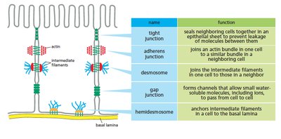
Classification of Cell Junctions
name | function |
|---|---|
tight junction | seals neighboring cells together in an epithelial sheet to prevent leakage of molecules between them |
adherens junction | joins an actin bundle in one cell to a similar bundle in a neighboring cell |
desmosome | joins the intermediate filaments in one cell to those in a neighboring cell |
gap junction | forms channels that allow small water-soluble molecules, including ions, to pass from cell to cell |
hemidesmosome | anchors intermediate filaments in a cell to the basal lamina |
Example: Gap junctions in heart muscle allow electrical signals to pass rapidly between cells, coordinating contraction.
Additional info: The diagram and table above visually reinforce the classification and function of cell junctions, directly supporting the explanation of cell-cell and cell-matrix interactions.
