 Back
Cellular Energy Metabolism: Glycolysis, Fermentation, and Aerobic Respiration
Study Guide - Smart Notes
 Tailored notes based on your materials, expanded with key definitions, examples, and context.
Glycolysis and Cellular Energy Metabolism
Overview of Metabolic Pathways
Metabolic pathways are series of chemical reactions occurring within a cell that lead to the synthesis (anabolic) or breakdown (catabolic) of molecules. These pathways are essential for maintaining cellular function and energy balance.
Anabolic pathways: Synthesize complex molecules from simpler ones, requiring energy input (endergonic), increasing molecular order, and causing a local decrease in entropy.
Catabolic pathways: Break down complex molecules into simpler ones, releasing energy (exergonic), decreasing molecular order, and increasing entropy.
Roles of catabolic pathways:
Release free energy used to perform cellular work.
Provide metabolites for biosynthetic processes.
Metabolism can be aerobic (with oxygen) or anaerobic (without oxygen).

Cellular Energy Storage Molecules
how do we store chemical energy:
Cells store chemical energy in several key molecules, which are used to drive endergonic reactions and cellular processes.
ATP (Adenosine Triphosphate): The primary energy currency of the cell.
GTP (Guanosine Triphosphate): Used in protein synthesis and signal transduction.
Acetyl CoA: Central intermediate in metabolism, especially in the Krebs cycle.
Creatine phosphate: Serves as a rapid source of high-energy phosphate in muscle cells.
Reduced coenzymes (e.g., NADH, FADH2): Store electrons for use in oxidative phosphorylation.
ATP Hydrolysis and Free Energy
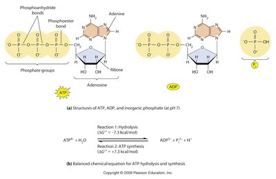
ATP hydrolysis is a key reaction that releases energy for cellular work. The hydrolysis of ATP to ADP and inorganic phosphate is highly exergonic.
Standard free energy change for ATP hydrolysis: kcal/mol.
ATP is regenerated from ADP and Pi during cellular respiration.
Standard Free Energies of Hydrolysis
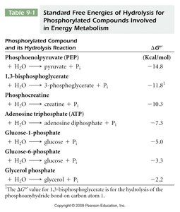
Different phosphorylated compounds have varying free energies of hydrolysis, which determines their role in metabolism.
Compound | Hydrolysis Reaction | (kcal/mol) |
|---|---|---|
Phosphoenolpyruvate (PEP) | PEP + H2O → pyruvate + Pi | -14.8 |
1,3-bisphosphoglycerate | 1,3-bisphosphoglycerate + H2O → 3-phosphoglycerate + Pi | -11.8 |
Phosphocreatine | Phosphocreatine + H2O → creatine + Pi | -10.3 |
ATP | ATP + H2O → ADP + Pi | -7.3 |
Glucose-1-phosphate | Glucose-1-phosphate + H2O → glucose + Pi | -5.0 |
Glucose-6-phosphate | Glucose-6-phosphate + H2O → glucose + Pi | -3.3 |
Glycerol phosphate | Glycerol phosphate + H2O → glycerol + Pi | -2.2 |
ATP as the Cellular Energy Currency
ATP is the most convenient and widely used energy currency in the cell, with a free energy of hydrolysis of -7.3 kcal/mol. It is used to power a variety of cellular processes, including biosynthesis, transport, and mechanical work.
Role of the ATP/ADP System
The ATP/ADP system acts as an energy buffer, allowing cells to rapidly respond to energy demands by interconverting ATP and ADP.
Oxidation and Reduction in Metabolism
Oxidation-reduction (redox) reactions are central to energy metabolism. Oxidation is exergonic (releases energy), while reduction is endergonic (stores energy in reduced compounds).
Oxidation: Loss of electrons or hydrogen atoms.
Reduction: Gain of electrons or hydrogen atoms.
Cells store energy by accumulating reduced compounds such as NADH and FADH2.
high energy bonds is how u store energy.
NAD+ and NADH: Electron Carriers
NAD (nicotinamide adenine dinucleotide) is a coenzyme derived from niacin (vitamin B3) and acts as an electron carrier in redox reactions, especially in glycolysis and the Krebs cycle.
NAD+ accepts electrons (is reduced) to form NADH.
NADH can donate electrons to the electron transport chain for ATP production.

Polymer Breakdown: Phosphorolytic Cleavage
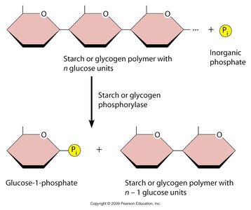
Polysaccharides such as glycogen and starch are broken down by phosphorolytic cleavage, where inorganic phosphate (Pi) breaks the bond between glucose units, producing glucose-1-phosphate, which can enter glycolysis.
Phosphorolytic cleavage is similar to hydrolysis but uses Pi instead of H2O.
Glucose-1-phosphate is converted to glucose-6-phosphate for glycolysis.
Glycolysis: Pathway and Regulation
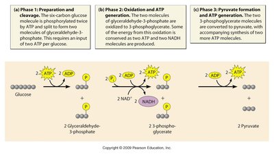
Glycolytic Pathway Overview be able to draw 6 carbons , 2 phosphate , you need 2 Atp molecules
Glycolysis is the metabolic pathway that converts glucose into pyruvate, generating ATP and NADH in the process. It occurs in the cytosol and does not require oxygen.
Net gain per glucose: 2 ATP, 2 NADH, and 2 pyruvate molecules.
Total free energy change: approximately -20 kcal/mol.
In the absence of oxygen, pyruvate is further metabolized by fermentation.
aerobic conditions to anaerobic , example : lactate in the muscles
Phases of Glycolysis
Phase 1: Preparation and cleavage – Glucose is phosphorylated and split into two three-carbon molecules (requires 2 ATP).
Phase 2: Oxidation and ATP generation – Glyceraldehyde-3-phosphate is oxidized, generating NADH and ATP.
Phase 3: Pyruvate formation and ATP generation – Further reactions yield pyruvate and additional ATP.

Fate of Pyruvate: Fermentation and Aerobic Respiration
After glycolysis, the fate of pyruvate depends on the presence or absence of oxygen.
Aerobic conditions: Pyruvate is converted to acetyl-CoA and enters the Krebs cycle for complete oxidation.
Anaerobic conditions: Pyruvate is reduced to lactate (in animals) or ethanol and CO2 (in yeast) via fermentation, regenerating NAD+ for glycolysis.

Gluconeogenesis and Regulation
Gluconeogenesis is the synthesis of glucose from non-carbohydrate precursors, primarily in the liver. Glycolysis and gluconeogenesis are reciprocally regulated, mainly through allosteric enzymes.
Key regulatory enzymes: hexokinase, phosphofructokinase, pyruvate kinase (glycolysis); fructose-1,6-bisphosphatase, PEP carboxykinase (gluconeogenesis).
Regulation is mostly allosteric, responding to cellular energy status.
Glycolysis breaks down glucose to produce energy, while gluconeogenesis synthesizes new glucose from non-carbohydrate sources. if we have glucose then glycolysis.

Aerobic Respiration and Mitochondrial Function
Overview of Aerobic Respiration
Aerobic respiration involves the flow of electrons from reduced coenzymes (NADH, FADH2) to a final electron acceptor (usually O2), resulting in the synthesis of ATP via oxidative phosphorylation.
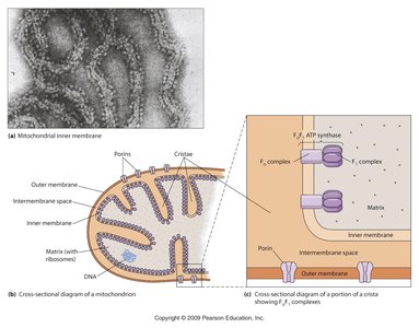
Electron transport chain (ETC) is located in the inner mitochondrial membrane.
Oxygen is the most common final electron acceptor, forming water.
Mitochondrial Structure and Function
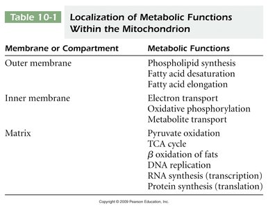
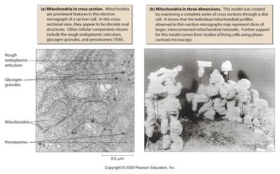
Mitochondria are the site of aerobic respiration and contain specialized compartments for different metabolic functions.
Membrane/Compartment | Metabolic Functions |
|---|---|
Outer membrane | Phospholipid synthesis, fatty acid desaturation and elongation |
Inner membrane | Electron transport, oxidative phosphorylation, metabolite transport |
Matrix | Pyruvate oxidation, TCA cycle, β-oxidation of fats, DNA/RNA synthesis, protein synthesis you need to know mitochondria 78% protein Ana krebs cycle |
Krebs Cycle (Citric Acid Cycle)
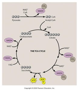
The Krebs cycle occurs in the mitochondrial matrix and completes the oxidation of acetyl-CoA to CO2, generating NADH, FADH2, and ATP (or GTP).
Each turn of the cycle yields: 3 NADH, 1 FADH2, 1 ATP (or GTP), and 2 CO2.
Key steps include de-carboxylation( losing carbon) and oxidation reactions.
Electron Transport Chain and ATP Synthase
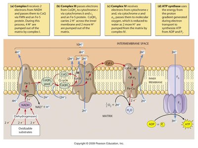
The electron transport chain (ETC) consists of four complexes that transfer electrons from NADH and FADH2 to oxygen, pumping protons across the inner mitochondrial membrane to generate a proton gradient.
Proton gradient drives ATP synthesis via ATP synthase (chemiosmotic coupling).
Q cycle allows additional protons to be pumped without additional electron transport.
how do we know if this is a domain/channel
ATP Synthase: Structure and Function
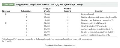
ATP synthase is a multi-subunit enzyme complex that synthesizes ATP from ADP and inorganic phosphate, powered by the flow of protons down their electrochemical gradient.
Structure | Polypeptide | Molecular Weight | Number Present | Function |
|---|---|---|---|---|
F0 | a | 30,000 | 1 | Proton channel |
F0 | b | 17,000 | 2 | Peripheral stalk |
F0 | c | 8,000 | 10 | Rotating ring |
F1 | α | 55,000 | 3 | Promotes activity |
F1 | β | 52,000 | 3 | Catalytic site |
F1 | γ | 36,000 | 1 | Stalk, energy transfer |
F1 | δ | 15,000 | 1 | Anchors F1 to F0 |
F1 | ε | 15,000 | 1 | Anchors γ subunit |
-- you need how to remember how much from what you get in these stages. glucose gives you ?
Brown Fat and Thermogenin
Brown adipose tissue contains the uncoupling protein thermogenin, which allows protons to re-enter the mitochondrial matrix without generating ATP, releasing energy as heat. This process is important for thermoregulation in newborn mammals and hibernating animals.
Uncoupling ATP synthesis from electron transport increases heat production.
Brown fat is more abundant in hibernating animals and newborns.
If all mitochondria contained thermogenin, ATP production would be severely impaired, leading to energy deficiency.
Additional info: The notes above integrate and expand upon the provided content, ensuring a comprehensive and self-contained study guide for college-level cell biology students. All images included are directly relevant to the adjacent explanations and reinforce key concepts in cellular energy metabolism.

brown fat slide. do you expect to have more brown fat than a bear in hibernation
