 Back
BackCellular Movement: Motility and Contractility
Study Guide - Smart Notes
 Tailored notes based on your materials, expanded with key definitions, examples, and context.
Cellular Movement: Motility and Contractility
Introduction to Cellular Motility
Cellular movement, or motility, is a fundamental property of living cells, enabling them to move themselves, their environment, or intracellular components. Contractility is a specialized form of motility, most notably observed in muscle cells, where it results in cell shortening and force generation.
Motile Systems in Eukaryotic Cells
Levels of Motility
Tissue Level: Coordinated movement of groups of cells (e.g., muscle contraction).
Cellular Level: Movement of individual cells (e.g., sperm swimming, white blood cell migration).
Subcellular Level: Movement of organelles and vesicles within cells (e.g., chromosome separation during mitosis).
Motility is driven by the cytoskeleton, primarily microtubules (MTs) and microfilaments (MFs), which serve as tracks for motor proteins that convert chemical energy into mechanical work.
Two Eukaryotic Motility Systems
Microtubule-Based Motility
Microtubule-based motility is essential for processes such as axonal transport in neurons and the movement of cilia and flagella. The primary motor proteins involved are kinesins and dyneins.
Kinesins: Move cargo toward the plus end of microtubules (anterograde transport).
Dyneins: Move cargo toward the minus end of microtubules (retrograde transport).
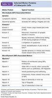
Table: Selected Motor Proteins of Eukaryotic Cells
Microfilament-Based Motility
Microfilament-based motility is exemplified by muscle contraction and other actin-dependent processes. The main motor proteins are myosins, which generally move toward the plus end of actin filaments.
Myosin II: Responsible for muscle contraction.
Other myosins: Involved in vesicle transport, cell movement, and cytokinesis.
Molecular Motors: Structure and Function
Common Features of Motor Proteins
All motor proteins couple ATP hydrolysis to conformational changes that generate movement.
They undergo cycles of ATP binding, hydrolysis, and product release, resulting in processive movement along cytoskeletal filaments.
They have distinct domains for filament binding, cargo binding, and ATP hydrolysis.
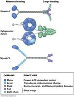
Figure: Domain organization of kinesin, dynein, and myosin V
Microtubule-Based Movement: Kinesins and Dyneins
Axonal Transport
Proteins and organelles are transported along axons by kinesins (anterograde) and dyneins (retrograde). This process is essential for neuronal function and survival.
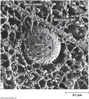
Example: Vesicle transport along microtubules in neurons
Kinesin Structure and Movement
Kinesin I consists of two heavy chains and two light chains.
Heavy chains have globular heads (bind MTs and ATP), a coiled-coil stalk, and a tail (binds cargo via light chains).
Kinesin moves in a "walking" manner, with each head alternately binding and stepping along the microtubule.
Processivity allows kinesin to move long distances without detaching.
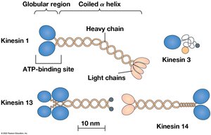
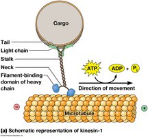
Figure: Kinesin structure and movement mechanism
Dynein Structure and Function
Dyneins are large, multi-subunit proteins found in the cytosol and axonemes (cilia/flagella).
Cytoplasmic dynein associates with dynactin to link to cargo.
Axonemal dyneins drive the sliding of microtubules in cilia and flagella.
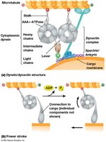
Figure: Dynein structure and mechanism
Microtubule-Based Motility: Cilia and Flagella
Structure and Function
Cilia and flagella are motile appendages with a common internal structure called the axoneme. Cilia are short and numerous, moving with an oarlike beat, while flagella are longer and fewer, moving with a wave-like motion.
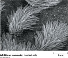
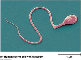
Examples: Cilia on tracheal cells and flagellum of a sperm cell
Axoneme and Basal Body Structure
The axoneme has a "9 + 2" arrangement: nine outer doublet microtubules and two central singlets.
Basal bodies anchor cilia/flagella and resemble centrioles (nine triplet microtubules).
Primary cilia have a "9 + 0" structure and are non-motile, serving sensory functions.
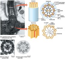
Figure: Axoneme and basal body structure
Mechanism of Bending
Axonemal dynein arms generate sliding between adjacent microtubule doublets.
Interdoublet links (nexin) and radial spokes convert sliding into bending.
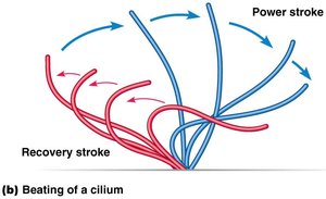
Figure: Sliding and bending mechanism in cilia and flagella
Beating Patterns
Cilia beat with a power stroke and recovery stroke, moving fluid parallel to the cell surface.
Flagella generate a propagated wave, moving the cell through fluid.
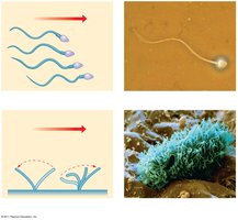
Figure: Beating patterns of cilia and flagella
Microfilament-Based Motility: Muscle Contraction
Muscle Types and Structure
Muscle contraction is the most familiar example of microfilament-based motility. There are three muscle types: skeletal (voluntary), cardiac (heart), and smooth (involuntary).
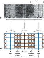
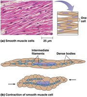
Figure: Skeletal and smooth muscle cell structure
Myosin Structure and Function
Myosins are ATP-dependent motors that move along actin filaments.
All myosins have a globular head (binds actin and ATP) and a tail (varies in length and function).
Type II myosins form thick filaments in muscle and mediate contraction by pulling actin filaments together.
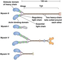
Figure: Myosin II, I, V, and VI structures
Muscle Cell Organization
Muscle fibers contain myofibrils, which are divided into repeating units called sarcomeres.
Sarcomeres contain thin (actin, troponin, tropomyosin) and thick (myosin) filaments.
Filaments are aligned, creating a pattern of dark (A bands) and light (I bands) regions.
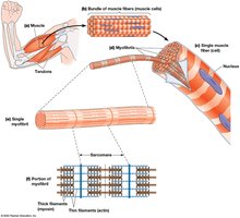
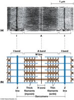
Figure: Sarcomere structure and banding pattern
Sliding-Filament Model of Contraction
Muscle contraction occurs as thin filaments slide past thick filaments, shortening the sarcomere without changing filament length. Myosin heads form transient cross-bridges with actin, powered by ATP hydrolysis.
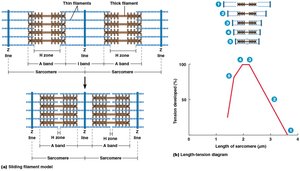
Figure: Sliding-filament model of muscle contraction
The Contraction Cycle
Myosin binds actin in a high-energy state (ADP + Pi bound).
Release of Pi triggers the power stroke, moving the thick filament toward the Z line.
ATP binding causes myosin to detach from actin; hydrolysis resets the head for another cycle.
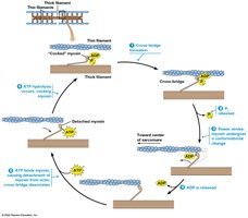
Figure: The contraction cycle in muscle
Regulation of Muscle Contraction by Calcium
At low Ca2+, tropomyosin blocks myosin-binding sites on actin.
At high Ca2+, Ca2+ binds troponin C, shifting tropomyosin and exposing binding sites.
Muscle contraction is initiated by nerve impulses that trigger Ca2+ release from the sarcoplasmic reticulum.
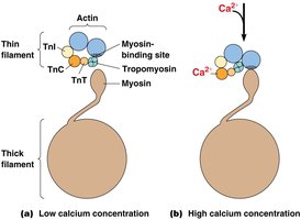
Figure: Calcium-dependent regulation of muscle contraction
Cardiac and Smooth Muscle
Cardiac muscle: Cells are joined by intercalated discs with gap junctions for electrical coupling; contraction is coordinated by pacemaker cells.
Smooth muscle: Cells are long, thin, and non-striated; contraction is slower and regulated by Ca2+-calmodulin activation of myosin light-chain kinase (MLCK).
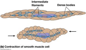
Figure: Structure and contraction of smooth muscle cells
Regulation of Smooth Muscle Contraction
Increased Ca2+ binds calmodulin, activating MLCK.
MLCK phosphorylates myosin light chains, enabling cross-bridge cycling with actin.
Relaxation occurs when Ca2+ levels fall and myosin light-chain phosphatase removes the phosphate.
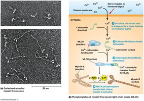
Figure: Regulation of smooth muscle contraction by Ca2+ and MLCK
Microfilament-Based Motility in Nonmuscle Cells
Cell Migration and Amoeboid Movement
Nonmuscle cells move by extending lamellipodia or filopodia, attaching to the substrate via integrins, and generating tension to pull the cell forward.
Amoeboid movement involves cycles of actin gelation (solidification) and solation (liquefaction) to form pseudopodia.
Cytoplasmic Streaming
Actomyosin-dependent movement of cytoplasm, known as cytoplasmic streaming or cyclosis in plants, helps distribute nutrients and organelles within large cells.
Summary Table: Selected Motor Proteins of Eukaryotic Cells
Motor Protein | Typical Function |
|---|---|
Cytoplasmic dynein | Moves cargo toward minus ends of MTs |
Axonemal dynein | Activates MT sliding in flagella and cilia |
Kinesin 1 | Direct, moves cargo toward plus ends of MTs |
Kinesin 2 | Direct, movement of organelles within cilia and flagella |
Kinesin 5 | Bidirectional, sliding of antiparallel MTs in mitosis |
Kinesin 13 | Depolymerizes MT ends |
Myosin II | Muscle contraction, cytokinesis |
Myosin V | Organelle and vesicle transport |
Myosin I | Membrane association, endocytosis |
Table: Main functions of selected motor proteins
Additional info: This guide covers the core concepts of cellular motility and contractility, including the molecular mechanisms, structural organization, and regulation of movement in eukaryotic cells. It integrates microtubule- and microfilament-based systems, highlighting their roles in both muscle and nonmuscle cells.
