 Back
BackCellular Movement: Motility and Contractility – Study Notes
Study Guide - Smart Notes
 Tailored notes based on your materials, expanded with key definitions, examples, and context.
Cellular Movement: Motility and Contractility
Overview of Cell Motility and Contractility
Cellular movement is a fundamental process in cell biology, encompassing the movement of cells, their environment, and intracellular components. Motility refers to the ability of cells or organisms to move, while contractility describes the shortening of muscle cells, a specialized form of motility. These processes are essential for development, immune responses, and tissue maintenance.
Cell motility includes movement through the environment, movement of the environment past or through a cell, and movement of components within the cell.
Contractility is primarily seen in muscle cells and involves the shortening of the cell.
Motility occurs at tissue, cellular, and subcellular levels.
Intracellular motility is crucial for processes like chromosome separation during cell division.
Motile Systems and the Cytoskeleton
Cellular movement is driven by the cytoskeleton, which provides a scaffold for motor proteins. The two main motility systems in eukaryotic cells are microtubule-based and microfilament-based motility.
Microtubule-based motility: Involves movement along microtubules, such as fast axonal transport in neurons and the sliding of microtubules in cilia and flagella.
Microfilament-based motility: Involves movement along actin filaments, such as muscle contraction.
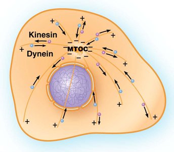
Microtubule-Based Movement: Kinesins and Dyneins
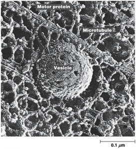
Microtubule-based motility relies on motor proteins, primarily kinesins and dyneins, which transport cellular cargo along microtubule tracks.
Kinesin I: Responsible for ATP-dependent transport toward the plus ends of microtubules (anterograde transport).
Cytoplasmic dynein: Moves cargo toward the minus ends of microtubules (retrograde transport).
Both proteins are essential for fast axonal transport, moving vesicles at rates of ~2 μm/sec.
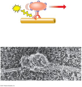
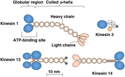
Kinesin Structure and Movement
Kinesins are complex proteins with distinct structural domains that enable their function as molecular motors.
Kinesins consist of two dimerized heavy chains and two light chains.
The heavy chains contain globular domains for microtubule and ATP binding, a coiled-coil stalk, and a lever-like neck.
The light chains bind to cargo.
Kinesin movement resembles "walking," with the two globular head domains alternating steps (~8 nm per step).
Kinesins exhibit processivity, allowing them to move long distances before detaching.
Example: Fast axonal transport involves kinesin moving vesicles at a rate of about 2 μm/sec, requiring approximately 250 steps per second (since each step is 8 nm).
Dyneins and Their Functions
Dyneins are another class of motor proteins, found in both the cytosol and axonemes. They are (-) end-directed and ATP-dependent.
Two types of cytoplasmic dynein are identified, associating with the dynactin complex for cargo binding.
Axonemal dyneins are involved in cilia and flagella movement.
All dyneins move toward the minus end of microtubules.
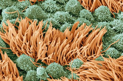
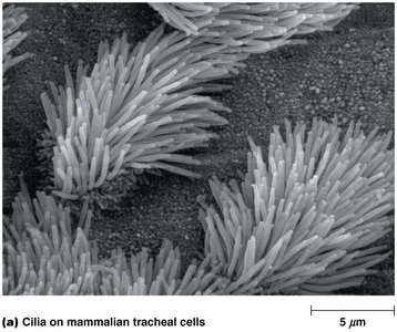
Microtubule-Based Motility: Cilia and Flagella
Cilia and flagella are motile appendages of eukaryotic cells, sharing a common structural basis. Microtubules are essential for their movement.
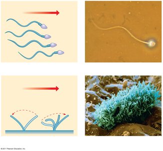
Cilia: Short (2–10 μm), numerous, and display an oar-like beating pattern generating force parallel to the cell surface.
Flagella: Longer (up to 200 μm), fewer per cell, and move with a propagated bending motion generating force parallel to the flagellum.
Structure of Cilia and Flagella
Cilia and flagella consist of an axoneme connected to a basal body, surrounded by an extension of the cell membrane. The axoneme has a characteristic "9 + 2" pattern: 9 outer doublets and 2 central microtubules.
The basal body resembles a centriole, with nine sets of microtubule triplets.
Primary cilia, used in sensory structures, have a "9 + 0" structure and are non-motile.
The central pair of microtubules is required for motility.
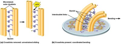
Axonemal Dynein and Doublet Sliding
Axonemal dynein mediates the sliding of microtubule doublets within the axoneme, causing bending of cilia and flagella. Interdoublet links, such as nexin, limit the extent of movement and convert sliding into bending.
Dynein forms links between microtubule doublets, moving along one doublet while the other serves as cargo.
Interdoublet links and crosslinks are responsible for coordinated bending.
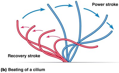
Beating Patterns of Cilia and Flagella
Cilia and flagella exhibit distinct beating patterns that propel cells or move fluid across cell surfaces.
Cilia display a power stroke and recovery stroke, generating movement parallel to the cell surface.
Flagella move with a propagated bending motion, propelling cells through fluid.
Intraflagellar Transport (IFT)
Intraflagellar transport is the process by which tubulin subunits and other components are shuttled to and from the growing tips of flagella and cilia. Both plus- and minus-end-directed motor proteins are involved.
Kinesins move material to the tips of flagella.
Dynein brings material back toward the base.
Summary Table: Comparison of Cilia and Flagella
Feature | Cilia | Flagella |
|---|---|---|
Length | 2–10 μm | Up to 200 μm |
Number per cell | Many | One or few |
Beating pattern | Oar-like, power/recovery stroke | Propagated bending motion |
Structural pattern | 9 + 2 axoneme | 9 + 2 axoneme |
Function | Move fluid across cell surface | Propel cell through fluid |
Additional info: The central pair of microtubules in the axoneme is essential for motility, as indicated by the difference between motile cilia (9 + 2) and non-motile primary cilia (9 + 0).
