 Back
BackChapter 1: Cells – The Fundamental Units of Life (Mini-Textbook Study Notes)
Study Guide - Smart Notes
 Tailored notes based on your materials, expanded with key definitions, examples, and context.
Cells: The Fundamental Units of Life
Unity and Diversity of Cells
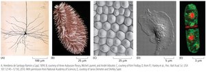
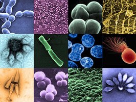
Cells are the basic structural and functional units of all living organisms. They exhibit remarkable diversity in size, shape, and function, yet share fundamental biochemical processes.
Cell Diversity: Cells can be specialized for various functions, such as neurons for signaling, macrophages for immunity, yeast for reproduction, and plant cells for photosynthesis.
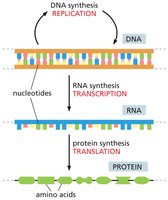
Basic Chemistry: All cells follow the Central Dogma of molecular biology: DNA is replicated, transcribed into RNA, and translated into protein.
Self-Replication: Cells possess the ability to replicate themselves, requiring organization, genetic blueprints, materials, energy, and catalytic activity.
Feedback Loops: Cellular processes are regulated by feedback mechanisms to maintain homeostasis.
Cell Theory: All living cells arise from the growth and division of pre-existing cells, a principle established through repeated scientific observation.
Evolution: All cells are believed to have evolved from a common ancestral cell, with mutations in the genome driving evolutionary adaptation.
Differentiation: Cells in multicellular organisms differentiate by activating or silencing specific genes, despite sharing the same genome.
Microscopy of Cells
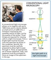
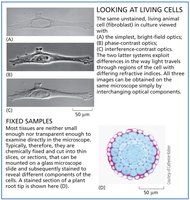
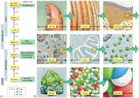
Microscopy is essential for studying cells, allowing visualization of structures at various scales. Different types of microscopy provide unique advantages in resolution and contrast.
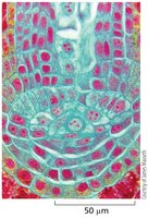
Conventional Light Microscopy: Uses visible light and glass lenses to magnify cells up to 1000X, with a resolution limit of 0.2 μm.
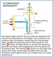
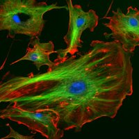
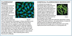
Fluorescence Light Microscopy: Utilizes fluorescent dyes and filters to visualize specific cell components with high contrast.
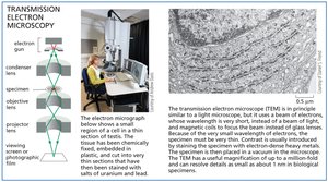
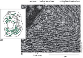
Transmission Electron Microscopy (TEM): Employs electron beams and heavy metal stains to achieve magnification up to 1,000,000X and resolution down to 1 nm.
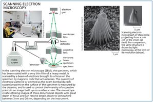
Scanning Electron Microscopy (SEM): Provides detailed surface images with resolution between 3–20 nm.
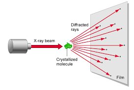
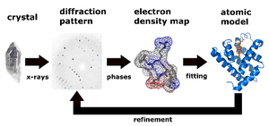
Nanoscopy/X-ray Crystallography: Allows determination of atomic positions in biomolecules, with resolution as fine as 0.15 nm.
Prokaryotic Cells
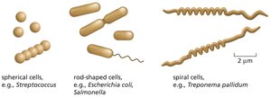
Prokaryotes are the most numerous and diverse cells on Earth, characterized by their simplicity and lack of a nucleus. They are divided into two domains: Bacteria and Archaea.
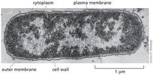
Structure: Prokaryotic cells are typically small (a few μm), with essential components for life.
No Nucleus: Their genome is not enclosed in a membrane-bound nucleus.
Bacteria vs. Archaea: While similar in appearance, these domains differ significantly in DNA sequence and environmental adaptations.
Examples: E. coli (Bacteria), Archaea (found in extreme environments).

Eukaryotic Cells
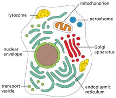
Eukaryotic cells are distinguished by their compartmentalization, including a nucleus and various organelles. These features enable complex functions and specialization.
Nucleus: Stores genetic information within a double membrane.
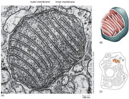
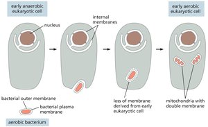
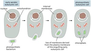
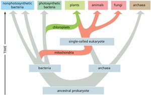
Mitochondria: Generate energy (ATP) from food molecules, contain their own DNA, and are believed to have evolved from engulfed bacteria.
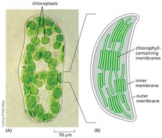
Chloroplasts: Capture energy from sunlight for photosynthesis, also contain their own DNA, and likely evolved from engulfed photosynthetic bacteria.
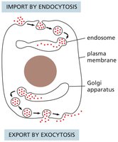
Internal Membranes: Create compartments such as the endoplasmic reticulum (ER), Golgi apparatus, lysosomes, and peroxisomes, each with specialized functions.
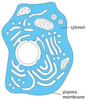
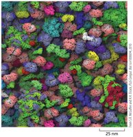
Cytosol: The aqueous gel-like substance filling the cell, rich in proteins and metabolites.
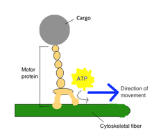
Cytoskeleton: Provides structural support, shape, and facilitates intracellular transport.

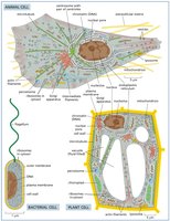
Cellular Architecture
Cellular architecture refers to the spatial organization of cellular components, which is crucial for proper function and interaction.
Scale Bars: Used in microscopy to indicate the size of structures.
Comparison: Animal, plant, and bacterial cells have distinct architectures reflecting their functions.
Origin of Life and Cells
The origin of life is traced back to approximately 3.5 billion years ago, with the emergence of the first cells. Eukaryotic cells appeared later, around 1.8 billion years ago.
Timeline: Evolution from simple prokaryotes to complex eukaryotes.
Phylogeny: Modern cells share ancestry with ancient prokaryotes.
Model Organisms
Model organisms are species extensively studied to understand fundamental biological processes. They provide insights applicable to other organisms, including humans.
Escherichia coli: Prokaryote, easy to grow, rapid reproduction, fundamental processes similar to human cells.
Saccharomyces cerevisiae: Eukaryotic yeast, rapid reproduction, used to study internal compartments.
Arabidopsis thaliana: Model plant, short generation time, genes relevant to agriculture.
Drosophila melanogaster: Fruit fly, genetic studies, developmental genes similar to humans.
Caenorhabditis elegans: Nematode, genome sequenced, developmental studies, many human gene counterparts.
Zebrafish: Vertebrate, transparent embryos, direct observation of development.
Mice: Mammals, genetic studies, engineered mutations, similar genes to humans.
Human Cells: Used to study differentiation and specialized functions.

Genomes
Genomes vary in size across species but often encode similar genes, reflecting evolutionary conservation and functional similarities.
Genome: The complete set of DNA in an organism.
Gene Conservation: Many genes are shared across diverse species, enabling comparative studies.
