 Back
BackCH 7 - Membranes – Structure, Function, and Chemistry
Study Guide - Smart Notes
 Tailored notes based on your materials, expanded with key definitions, examples, and context.
Membranes: Structure, Function, and Chemistry
Overview of Membrane Functions
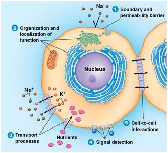
Biological membranes are essential for defining the boundaries of cells and their internal compartments. They play multiple roles in cellular life, including compartmentalization, selective permeability, signal detection, and cell-to-cell interactions.
Boundary and Permeability Barriers: Membranes separate the cell from its environment and compartmentalize internal structures, creating distinct microenvironments.
Organization and Localization of Function: Specific cellular functions are associated with particular membranes due to the localization of functional molecules.
Transport Processes: Membrane proteins regulate the movement of substances across the membrane, either by direct diffusion or via specific transporters.
Signal Detection: Membranes contain receptors that detect chemical and electrical signals from the environment.
Cell-to-Cell Adhesion and Communication: Membranes mediate adhesion and communication between cells, critical for multicellular organization.
Boundary and Permeability Barriers
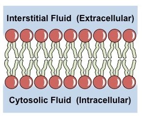
The plasma membrane surrounds the entire cell, while intracellular membranes compartmentalize functions within organelles. The hydrophobic interior of the membrane acts as an effective barrier to most polar molecules.
Organization and Localization of Function
Membranes are associated with specific cellular functions because the molecules responsible for these functions are embedded in or localized on the membranes. This organization allows for efficient metabolic processes and signaling pathways.
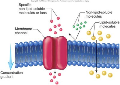
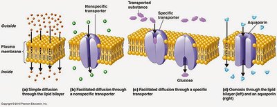
Transport Processes Across Membranes
Transport across membranes is mediated by proteins that facilitate the movement of ions and molecules. Some substances diffuse directly through the lipid bilayer, while others require specific transporters or channels.
Simple Diffusion: Movement of small, nonpolar molecules directly through the lipid bilayer.
Facilitated Diffusion: Movement of specific molecules via membrane proteins (channels or carriers).
Osmosis: Diffusion of water across the membrane, often through aquaporins.
Signal Detection
Cells receive information from their environment through chemical or electrical signals. Chemical signals bind to membrane proteins called receptors, while electrical signals can pass through channel proteins such as gap junctions.
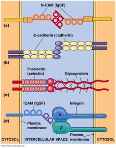
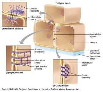
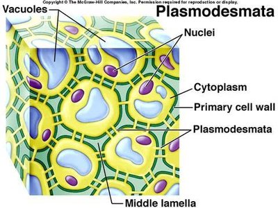
Cell-to-Cell Adhesion and Communication
Cell adhesion is critical for the development and maintenance of multicellular organisms. Cadherins are a major class of adhesion molecules that require calcium ions for function and mediate adhesion between similar cell types. Other junctions include tight junctions, gap junctions (in animals), and plasmodesmata (in plants).
Adhesive Junctions: Hold cells together.
Tight Junctions: Form seals that block passage of fluids between cells.
Gap Junctions: Allow rapid communication between adjacent cells.
Plasmodesmata: Enable intercellular communication in plants.
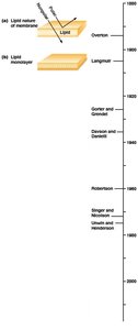
Models of Membrane Structure: Historical Perspective
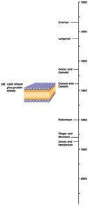
Development of Membrane Models
The understanding of membrane structure has evolved through experimental observations:
1890s (Overton): Proposed a lipid "coat" at the cell surface based on the permeability of lipid-soluble substances.
1900s (Langmuir): Demonstrated that phospholipids are amphipathic and orient with hydrophobic tails away from water.
1925 (Gorter and Grendel): Proposed the lipid bilayer model after extracting lipids from red blood cells.
1935 (Davson and Danielli): Suggested a protein-lipid-protein "sandwich" model to explain membrane properties.
1960s (Robertson): Electron microscopy revealed the trilaminar structure of membranes (unit membrane).
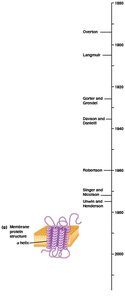
1972 (Singer and Nicholson): Introduced the fluid mosaic model, describing membranes as a fluid lipid bilayer with a mosaic of proteins.
Major Shortcomings of Early Models
The Davson–Danielli model could not explain the chemical diversity of membranes, the protein/lipid ratio, or the susceptibility of membranes to phospholipases. The fluid mosaic model resolved these inconsistencies by proposing that proteins are embedded within, not just coating, the lipid bilayer.
Membrane Composition and Microdomains
Lipid Components of Membranes
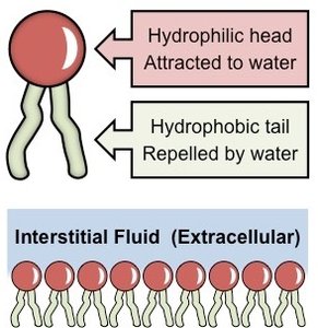
Membranes are primarily composed of lipids and proteins. The main classes of membrane lipids are phospholipids, glycolipids, and sterols.
Phospholipids: Most abundant, can be glycerol-based (phosphoglycerides) or sphingosine-based (sphingolipids).
Glycolipids: Lipids with carbohydrate groups, important in cell recognition.
Sterols: Cholesterol (animals), phytosterols (plants), and ergosterol (fungi) stabilize membranes and modulate fluidity.
Fatty Acids and Membrane Fluidity
Fatty acids are essential for membrane structure and function. Their length and degree of saturation influence membrane fluidity and the transition temperature ().
Saturated fatty acids: No double bonds, pack tightly, higher , less fluid.
Unsaturated fatty acids: One or more double bonds (usually cis), introduce kinks, lower , more fluid.
Sterols: Act as fluidity buffers, decreasing fluidity at high temperatures and preventing tight packing at low temperatures.
Membrane Asymmetry
Membrane lipids are distributed asymmetrically between the two monolayers. Glycolipids are typically found in the outer layer, and the degree of fatty acid saturation can differ between layers. Asymmetry is established during membrane synthesis and is maintained over time.
Lipid Rafts and Microdomains
Membranes are not homogenous; they contain dynamic microdomains called lipid rafts, which are rich in cholesterol and glycosphingolipids. These rafts influence membrane fluidity, protein trafficking, and signal transduction.
Membrane Proteins: The "Mosaic" Part of the Model
Classes of Membrane Proteins
Membrane proteins are classified based on their association with the lipid bilayer:
Integral (Transmembrane) Proteins: Span the bilayer with hydrophobic segments; can be singlepass or multipass.
Peripheral Proteins: Bound to membrane surfaces by weak interactions; do not penetrate the bilayer.
Lipid-Anchored Proteins: Covalently attached to lipids embedded in the bilayer.
Isolation and Analysis of Membrane Proteins
Membrane proteins can be isolated using detergents (for integral proteins) or by altering pH/ionic strength (for peripheral and lipid-anchored proteins). Electrophoresis (e.g., SDS-PAGE) and Western blotting are used to separate and identify proteins. Affinity labeling and membrane reconstitution (using liposomes) are advanced techniques for studying protein function.
Glycosylation and Glycoproteins
Many membrane proteins are glycosylated, with carbohydrate chains covalently linked to amino acid side chains. Glycosylation occurs in the ER and Golgi and is important for cell-cell recognition, immunity, and blood group antigens. The glycocalyx is a carbohydrate-rich surface coat involved in cell recognition.
Mobility and Organization of Membrane Proteins
Membrane proteins vary in their mobility; some move freely, while others are anchored to cytoskeletal or extracellular structures. Protein aggregation and anchoring restrict mobility, creating functional domains such as lipid rafts. Experimental evidence (e.g., cell fusion and photobleaching recovery) supports these observations.
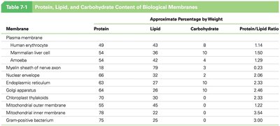
Table: Protein, Lipid, and Carbohydrate Content of Biological Membranes
Membrane | Protein (%) | Lipid (%) | Carbohydrate (%) | Protein/Lipid Ratio |
|---|---|---|---|---|
Plasma membrane | 49 | 43 | 8 | 1.14 |
Human erythrocyte | 49 | 43 | 8 | 1.14 |
Mammalian liver cell | 54 | 36 | 10 | 1.50 |
Myelin sheath of nerve axon | 18 | 79 | 3 | 0.23 |
Nuclear envelope | 66 | 32 | 2 | 2.06 |
Endoplasmic reticulum | 63 | 34 | 3 | 1.85 |
Golgi apparatus | 64 | 33 | 3 | 1.94 |
Chloroplast thylakoids | 70 | 30 | 0 | 2.33 |
Mitochondrial outer membrane | 59 | 41 | 0 | 1.44 |
Mitochondrial inner membrane | 76 | 24 | 0 | 3.17 |
Gram-positive bacterium | 75 | 25 | 0 | 3.00 |
Summary
Biological membranes are dynamic structures composed of lipids, proteins, and carbohydrates. Their fluid mosaic organization allows for compartmentalization, selective transport, signal transduction, and cell communication. The composition and structure of membranes are finely tuned to support the diverse functions required for cellular life.
