 Back
BackChemical Components of Cells: Amino Acids, Nucleic Acids, Sugars, and Lipids
Study Guide - Smart Notes
 Tailored notes based on your materials, expanded with key definitions, examples, and context.
Chemical Components of Cells
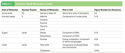
Common Small Molecules in Cells
Cells are composed of a variety of small molecules that serve as the building blocks for macromolecules. These include amino acids, aromatic bases, sugars, and lipids, each with distinct roles in cellular structure and function.
Kind of Molecules | Number Present | Names of Molecules | Role in Cell |
|---|---|---|---|
Amino acids | 20 | See list below | Monomeric units of all proteins |
Aromatic bases | 5 | Adenine, Cytosine, Guanine, Thymine, Uracil | Components of nucleic acids |
Sugars | varies | Ribose, Deoxyribose, Glucose | Components of RNA, DNA, energy metabolism |
Lipids | varies | Fatty acids, Cholesterol | Components of membranes, energy storage |
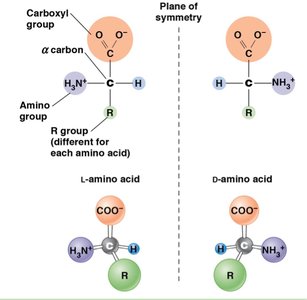
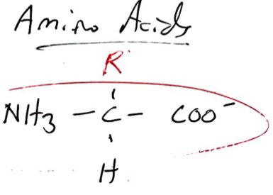
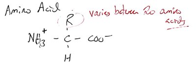
Amino Acids: Structure and Classification
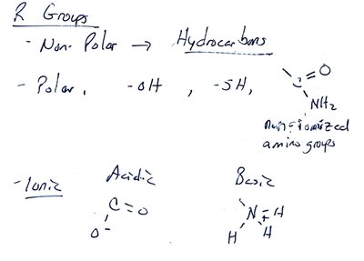
Amino acids are the monomeric units of proteins. Each amino acid has a central carbon (α carbon) bonded to an amino group, a carboxyl group, a hydrogen atom, and a variable R group. The R group determines the properties and classification of the amino acid.
Basic structure: NH3+–C–COO− with an R group and a hydrogen atom.
Isomerism: Amino acids exist as L and D isomers, but only L isomers are used in protein synthesis.
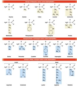
Classification: Based on the R group, amino acids can be nonpolar (hydrophobic), polar (hydrophilic), or charged (acidic/basic).
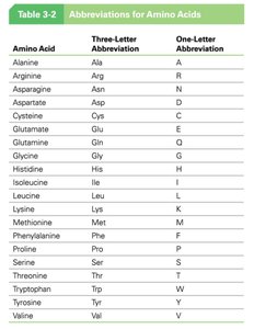
Amino Acid | Three-Letter Abbreviation | One-Letter Abbreviation |
|---|---|---|
Alanine | Ala | A |
Arginine | Arg | R |
Asparagine | Asn | N |
Aspartate | Asp | D |
Cysteine | Cys | C |
Glutamate | Glu | E |
Glutamine | Gln | Q |
Glycine | Gly | G |
Histidine | His | H |
Isoleucine | Ile | I |
Leucine | Leu | L |
Lysine | Lys | K |
Methionine | Met | M |
Phenylalanine | Phe | F |
Proline | Pro | P |
Serine | Ser | S |
Threonine | Thr | T |
Tryptophan | Trp | W |
Tyrosine | Tyr | Y |
Valine | Val | V |
Groups of Amino Acids
Nonpolar (hydrophobic): Glycine, Alanine, Valine, Leucine, Isoleucine, Methionine, Phenylalanine, Proline
Polar (hydrophilic): Serine, Threonine, Cysteine, Tyrosine, Asparagine, Glutamine
Charged: Acidic (Aspartate, Glutamate), Basic (Lysine, Arginine, Histidine)
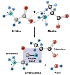
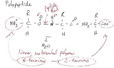
Peptide Bond Formation and Polypeptides
Amino acids are linked by peptide bonds formed through dehydration reactions. The resulting polypeptide is a linear, unbranched polymer with an N-terminus and a C-terminus.
Peptide bond: Covalent bond between the carboxyl group of one amino acid and the amino group of another.
Directionality: Polypeptides are synthesized from N-terminus to C-terminus.
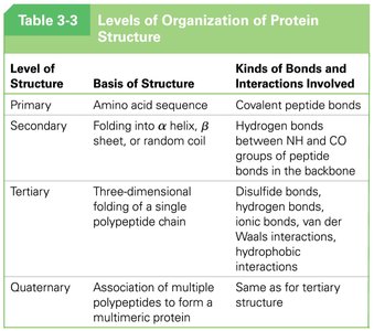
Levels of Protein Structure
Proteins exhibit four levels of structural organization: primary, secondary, tertiary, and quaternary. Each level is stabilized by specific types of bonds and interactions.
Level of Structure | Basis of Structure | Kinds of Bonds and Interactions Involved |
|---|---|---|
Primary | Amino acid sequence | Covalent peptide bonds |
Secondary | Folding into α helix, β sheet, or random coil | Hydrogen bonds between NH and CO groups |
Tertiary | Three-dimensional folding of a single polypeptide chain | Disulfide bonds, hydrogen bonds, ionic bonds, van der Waals, hydrophobic interactions |
Quaternary | Association of multiple polypeptides | Same as for tertiary structure |
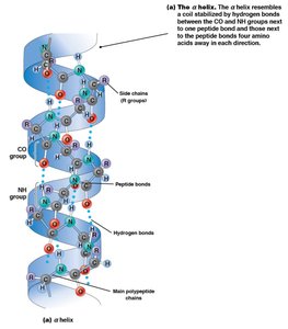
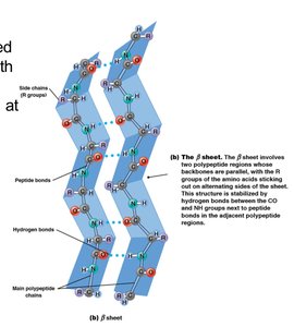
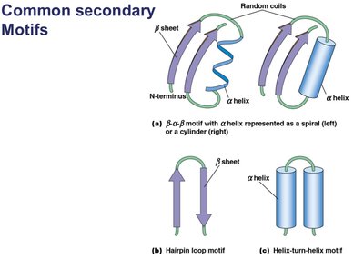
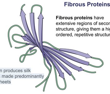
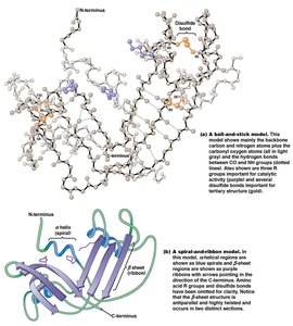
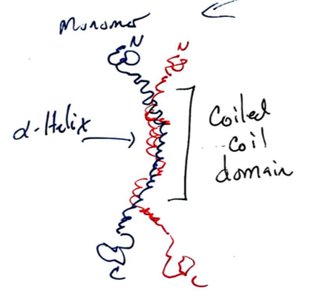
Secondary Structure: α Helix and β Sheet
α Helix: Spiral structure stabilized by hydrogen bonds between NH and CO groups four amino acids apart.
β Sheet: Sheet-like structure stabilized by hydrogen bonds between adjacent polypeptide regions; can be parallel or antiparallel.
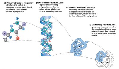
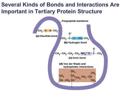
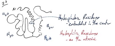
Tertiary Structure
Tertiary structure is the overall three-dimensional shape of a single polypeptide, determined by interactions among R groups.
Hydrophobic residues: Tend to be buried in the interior.
Hydrophilic residues: Tend to be exposed on the surface.
Stabilizing interactions: Disulfide bonds, hydrogen bonds, ionic bonds, van der Waals forces.
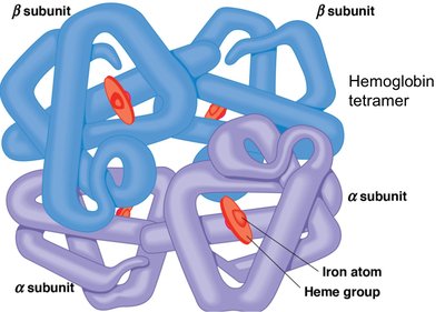
Quaternary Structure
Quaternary structure refers to the association of two or more polypeptide chains to form a functional protein complex. Hemoglobin is a classic example, consisting of two α and two β subunits.
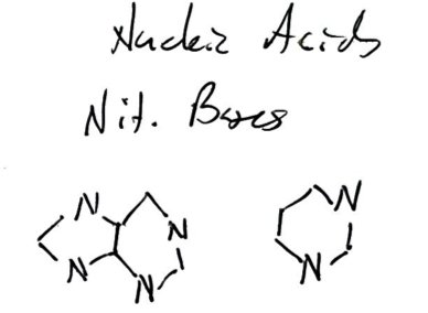
Nucleic Acids: Structure and Components
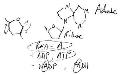
Nucleic acids (DNA and RNA) are polymers of nucleotides, each composed of a phosphate group, a five-carbon sugar (ribose or deoxyribose), and a nitrogenous base (purine or pyrimidine).
Purines: Adenine (A), Guanine (G)
Pyrimidines: Cytosine (C), Thymine (T, in DNA), Uracil (U, in RNA)
Nucleoside: Sugar + base
Nucleotide: Sugar + base + phosphate group
Bases | Nucleoside (RNA) | Nucleotide (RNA) | Deoxynucleoside (DNA) | Deoxynucleotide (DNA) |
|---|---|---|---|---|
Adenine (A) | Adenosine | Adenosine monophosphate (AMP) | Deoxyadenosine | Deoxyadenosine monophosphate (dAMP) |
Guanine (G) | Guanosine | Guanosine monophosphate (GMP) | Deoxyguanosine | Deoxyguanosine monophosphate (dGMP) |
Cytosine (C) | Cytidine | Cytidine monophosphate (CMP) | Deoxycytidine | Deoxycytidine monophosphate (dCMP) |
Uracil (U) | Uridine | Uridine monophosphate (UMP) | - | - |
Thymine (T) | - | - | Deoxythymidine | Deoxythymidine monophosphate (dTMP) |
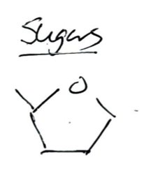
Sugars in Cells
Sugars are essential components of nucleic acids and serve as energy sources. Ribose is found in RNA, deoxyribose in DNA, and glucose is a primary energy source.
Lipids in Cells
Lipids are hydrophobic molecules that serve as energy storage and structural components of membranes. Fatty acids and cholesterol are key examples.
Fatty acids: Building blocks for other lipids
Cholesterol: Component of membranes
Summary Table: Levels of Organization of Protein Structure
Level of Structure | Basis of Structure | Kinds of Bonds and Interactions Involved |
|---|---|---|
Primary | Amino acid sequence | Covalent peptide bonds |
Secondary | Folding into α helix, β sheet, or random coil | Hydrogen bonds between NH and CO groups |
Tertiary | Three-dimensional folding of a single polypeptide chain | Disulfide bonds, hydrogen bonds, ionic bonds, van der Waals, hydrophobic interactions |
Quaternary | Association of multiple polypeptides | Same as for tertiary structure |
Key Points
Amino acids are the monomers of proteins, classified by their R groups.
Nucleic acids are polymers of nucleotides, which consist of a sugar, a phosphate, and a nitrogenous base.
Sugars are energy sources and structural components of nucleic acids.
Lipids are hydrophobic molecules important for energy storage and membrane structure.
