 Back
BackCytoskeletal Systems: Structure, Function, and Dynamics
Study Guide - Smart Notes
 Tailored notes based on your materials, expanded with key definitions, examples, and context.
Cytoskeletal Systems
Overview of the Cytoskeleton
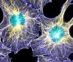
The cytoskeleton is a dynamic network of protein filaments that provides structural support, determines cell shape, and facilitates intracellular transport, cell division, and motility. It is composed of three major types of filaments: microtubules, microfilaments (actin filaments), and intermediate filaments. Each type has distinct structural and functional properties, but they work together to maintain cellular integrity and function.
Major Structural Elements of the Cytoskeleton
Microtubules (MTs): Hollow tubes (~25 nm diameter) made of α- and β-tubulin dimers; largest cytoskeletal element.
Microfilaments (Actin Filaments): Solid rods (~7 nm diameter) composed of actin monomers; smallest cytoskeletal element.
Intermediate Filaments (IFs): Rope-like fibers (~10 nm diameter) made from various proteins (e.g., keratins, vimentin, lamins); provide mechanical strength.
Microtubules
Structure and Assembly
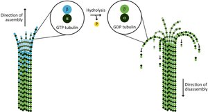
Microtubules are straight, hollow cylinders composed of 13 protofilaments, each made of αβ-tubulin heterodimers. They exhibit polarity, with a plus (+) end (fast-growing) and a minus (−) end (slow-growing). Microtubule assembly is a dynamic process involving nucleation, elongation, and plateau phases, regulated by GTP binding and hydrolysis.
Polymerization: Tubulin dimers add to the plus end in a GTP-bound state, forming a stabilizing "GTP cap."
Depolymerization: GTP hydrolysis to GDP destabilizes the microtubule, leading to shrinkage if the GTP cap is lost (dynamic instability).
Types and Functions of Microtubules
Cytoplasmic Microtubules: Maintain cell shape, form mitotic/meiotic spindles, position organelles, and direct vesicle transport.
Axonemal Microtubules: Found in cilia, flagella, and basal bodies; provide structure and motility to these organelles.
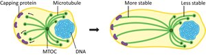
Microtubule-Organizing Centers (MTOCs) and Centrosomes
Microtubules originate from MTOCs, such as the centrosome in animal cells, which contains a pair of centrioles. The minus end is anchored at the MTOC, while the plus end extends outward.
Regulation of Microtubule Stability
Microtubule stability is regulated by various proteins and chemical agents:
Capping proteins: Stabilize microtubule ends, preventing depolymerization.
Microtubule-associated proteins (MAPs): Bind along microtubules to regulate their structure and interactions (e.g., Tau, MAP2).
Drugs: Colchicine, vinblastine, and nocodazole destabilize microtubules; paclitaxel (Taxol) stabilizes them. These are important in cancer therapy as antimitotic agents.
Dynamic Instability and Treadmilling
Microtubules undergo dynamic instability, alternating between phases of growth and shrinkage. Treadmilling occurs when the plus end grows while the minus end shrinks, resulting in a net movement of subunits through the filament.
Summary Table: Properties of Cytoskeletal Elements
Property | Microtubules | Microfilaments | Intermediate Filaments |
|---|---|---|---|
Diameter | ~25 nm | ~7 nm | ~10 nm |
Subunit | α/β-tubulin | Actin | Various (e.g., keratin, vimentin, lamin) |
Polarity | Yes | Yes | No |
Dynamic Instability | Yes | Yes | No |
Main Functions | Cell shape, transport, division | Motility, shape, contraction | Mechanical strength |
Microfilaments (Actin Filaments)
Structure and Assembly
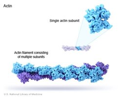
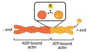
Microfilaments are composed of actin monomers (G-actin) that polymerize to form filamentous actin (F-actin). They are polar structures, with a fast-growing plus end and a slow-growing minus end. ATP binding and hydrolysis regulate their assembly and disassembly.
Functions of Microfilaments
Muscle contraction (with myosin)
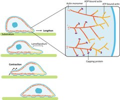
Cell migration and amoeboid movement
Cytokinesis (cell division)
Maintenance of cell shape and structure (especially in the cell cortex)
Vesicle and organelle transport
Regulation of Actin Filaments
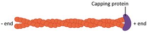
Capping proteins: Bind to filament ends to regulate growth and stability (e.g., CapZ at the plus end, tropomodulin at the minus end).
Crosslinking proteins: Organize actin into networks or bundles (e.g., filamin, α-actinin, fascin).
Severing proteins: Break filaments and cap new ends (e.g., gelsolin).
Specialized Actin Structures
Lamellipodia and Filopodia: Protrusive structures at the leading edge of crawling cells, formed by branched and bundled actin networks, respectively.
Microvilli: Finger-like projections in intestinal cells, supported by tightly bundled actin filaments, increasing surface area for absorption.
Red Blood Cell Cortex: Spectrin-ankyrin-actin network supports membrane flexibility and durability.
Intermediate Filaments
Structure and Assembly
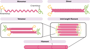
Intermediate filaments are rope-like fibers with a diameter between microtubules and microfilaments. They are composed of various proteins, classified into several types (e.g., keratins, vimentin, neurofilaments, nuclear lamins). IFs are assembled from coiled-coil dimers that form staggered tetramers, which then pack together to form the mature filament. Unlike MTs and actin filaments, IFs lack polarity and do not exhibit dynamic instability.
Functions of Intermediate Filaments
Provide mechanical strength to cells and tissues
Anchor organelles and maintain cell integrity
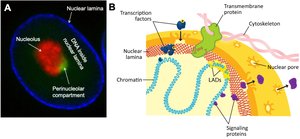
Support the nuclear envelope (nuclear lamins)
Resist tensile forces and deformation
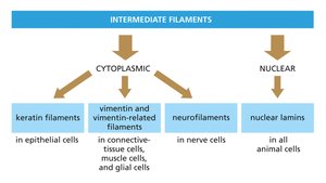
Examples of Intermediate Filament Proteins
Keratins: Found in epithelial cells, hair, and nails
Vimentin: Found in connective tissue, muscle, and glial cells
Neurofilaments: Found in neurons
Nuclear Lamins: Form a meshwork underlying the nuclear envelope in all animal cells

Integration of the Cytoskeleton
The cytoskeleton is a mechanically integrated structure. Microtubules resist compression, microfilaments generate tension, and intermediate filaments provide elasticity and resilience. These systems are interconnected, allowing cells to adapt to mechanical stress and maintain their architecture.
Summary Table: Chemical Agents Affecting the Cytoskeleton
Agent | Target | Effect | Application |
|---|---|---|---|
Colchicine | Microtubules | Destabilizes, prevents polymerization | Karyotyping, cancer therapy |
Vinblastine/Vincristine | Microtubules | Destabilizes, inhibits mitosis | Cancer therapy |
Nocodazole | Microtubules | Inhibits assembly (reversible) | Research, cancer therapy |
Paclitaxel (Taxol) | Microtubules | Stabilizes, prevents depolymerization | Cancer therapy |
Phalloidin | Actin filaments | Stabilizes filaments | Research |
Cytochalasin D | Actin filaments | Prevents polymerization | Research |
