Back
Cytoskeletal Systems: Structure, Function, and Dynamics
Study Guide - Smart Notes
 Tailored notes based on your materials, expanded with key definitions, examples, and context.
Cytoskeletal Systems
Overview of the Cytoskeleton
The cytoskeleton is a highly organized network of filaments and tubules within the cytosol of eukaryotic cells. It provides structural support, facilitates cell movement and division, and is dynamic, constantly assembling and disassembling in response to cellular needs.
Key Point 1: The cytoskeleton consists of interconnected filaments and tubules.
Key Point 2: It is essential for cell movement, division, and maintaining cell shape.
Example: The cytoskeleton enables processes such as mitosis, vesicle transport, and muscle contraction.
Major Structural Elements of the Cytoskeleton
Types of Cytoskeletal Elements
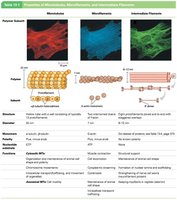
Eukaryotic cells possess three primary types of cytoskeletal elements: microtubules, microfilaments, and intermediate filaments. Each type has distinct structural and functional properties.
Microtubules: Composed of tubulin, ~25 nm diameter.
Microfilaments: Composed of actin, ~7 nm diameter.
Intermediate Filaments: Variable composition, 8–12 nm diameter.
Additional info: Septins are another class of polymer networks found in cells.
Bacterial Cytoskeletal Systems
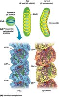
Bacteria and archaea possess cytoskeletal systems analogous to those in eukaryotes. These include actin-like and tubulin-like proteins that regulate cell shape and division.
MreB: Actin-like, involved in DNA segregation and cell shape.
FtsZ: Tubulin-like, regulates cell division.
Crescentin: Regulates cell shape.
Visualizing the Cytoskeleton
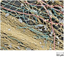
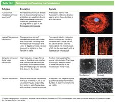
Various microscopic techniques are used to visualize cytoskeletal structures, including fluorescence microscopy and electron microscopy. These methods reveal the organization and dynamics of cytoskeletal elements.
Fluorescence microscopy: Uses fluorescent compounds to label cytoskeletal proteins.
Electron microscopy: Provides high-resolution images of filament networks.
Microtubules
Structure and Types of Microtubules
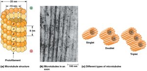
Microtubules are the largest cytoskeletal elements, forming straight, hollow cylinders. They are composed of α- and β-tubulin heterodimers arranged in protofilaments. Microtubules can exist as singlets, doublets, or triplets.
Singlet MTs: 13 protofilaments, found in cytoplasm.
Doublet and triplet MTs: Found in cilia, flagella, and basal bodies.
Axonemal MTs: Highly ordered bundles in cilia and flagella.
Microtubule Assembly and Dynamics
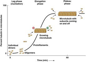
Microtubules assemble by the polymerization of tubulin dimers in the presence of GTP and Mg2+. The process involves nucleation (lag phase), elongation, and a plateau phase where assembly and disassembly are balanced.
Nucleation: Formation of oligomers as seeds for MT growth.
Elongation: Rapid addition of tubulin dimers.
Plateau: Equilibrium between assembly and disassembly.
Microtubule Treadmilling
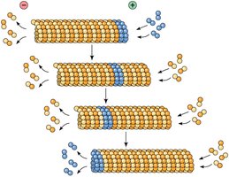
Treadmilling occurs when the concentration of free tubulin is above the critical concentration for the plus end but below that for the minus end, resulting in addition at the plus end and removal at the minus end.
Critical concentration: The minimum concentration of tubulin required for polymerization at each end.
Treadmilling: Dynamic instability of microtubules.
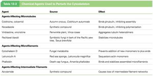
Drugs Affecting Microtubule Assembly
Certain chemicals can perturb microtubule function, affecting cell division and cytoskeletal stability. These agents are important in research and cancer therapy.
Colchicine: Binds tubulin, inhibits assembly, promotes disassembly.
Vinblastine/Vincristine: Antimitotic drugs, inhibit spindle assembly.
Microtubule-Organizing Centers (MTOCs)
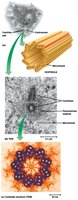
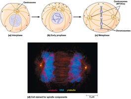
Microtubules originate from MTOCs, such as the centrosome in animal cells. The centrosome contains centrioles and pericentriolar material, and is crucial for organizing and polarizing microtubules.
Centrosome: Main MTOC in animal cells.
Centrioles: Nine pairs of triplet microtubules, involved in basal body formation.
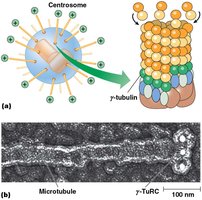
γ-tubulin: Found in centrosomes, nucleates MT assembly.
MTOCs Organize and Polarize Microtubules
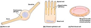
MTOCs anchor the minus ends of microtubules, allowing dynamic growth and shrinkage at the plus ends near the cell periphery. This organization is essential for cell polarity and division.
Polarity: Minus ends anchored, plus ends dynamic.
Cell division: MTOCs organize spindle fibers during mitosis.
Microtubule-Binding Proteins
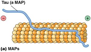
Microtubule-binding proteins regulate MT stability, transport, and structure. Some use ATP to drive transport, while others stabilize or destabilize MTs.
MAPs (Microtubule-Associated Proteins): Regulate MT structure and function.
Tau protein: Example of a MAP involved in stabilizing MTs.
Microfilaments
Structure and Function of Microfilaments
Microfilaments are the smallest cytoskeletal filaments, primarily composed of actin. They play key roles in muscle contraction, cell migration, and maintenance of cell shape.
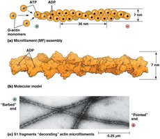
Actin: Protein building block, exists as G-actin (monomer) and F-actin (filament).
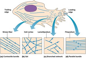
Cell cortex: Microfilaments just beneath the plasma membrane.
Microvilli: Structural core composed of actin bundles.
Polarity and Dynamics of Microfilaments
Microfilaments exhibit polarity, with rapid addition or loss of actin monomers at the plus end. ATP-actin is found at growing ends, while most of the filament is composed of ADP-actin.
Polarity: Plus end (barbed), minus end (pointed).
ATP hydrolysis: Drives filament dynamics.
Drugs Affecting Microfilament Polymerization
Specific drugs can modulate actin dynamics, affecting cell movement and structure.
Cytochalasins: Prevent addition of new monomers.
Latrunculin A: Sequesters actin monomers.
Phalloidin: Stabilizes microfilaments.
Microfilament Arrangements in Cells
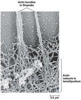
Actin filaments are organized into various structures depending on cell type and function, including stress fibers, cell cortex gels, lamellipodia, and filopodia.
Stress fibers: Contractile bundles in adherent cells.
Lamellipodia: Branched networks at the leading edge of crawling cells.
Filopodia: Parallel bundles for cell movement.
Actin-Binding Proteins
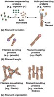
Actin-binding proteins regulate the polymerization, length, and organization of actin filaments, allowing precise control of cytoskeletal architecture.
Nucleation: Initiation of filament formation.
Elongation: Growth of filaments.
Severing and capping: Control filament length.
Crosslinking and bundling: Organize filaments into networks or bundles.
Microvilli Structure
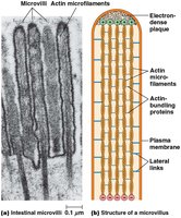
Microvilli are finger-like projections on the surface of intestinal mucosal cells, increasing surface area for absorption. Their core consists of tightly bundled actin filaments.
Actin bundles: Provide structural support.
Microvilli: Enhance cell surface area.
Intermediate Filaments
Structure and Properties of Intermediate Filaments
Intermediate filaments are abundant in animal cells and provide mechanical strength. They are the most stable and least soluble cytoskeletal components, supporting the entire cytoskeleton.
Keratin: Major IF in skin and related structures.
Stability: IFs are resistant to chemical attack.
Tissue Specificity and Classes of Intermediate Filaments
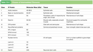
IF proteins vary greatly in composition and are grouped into six classes, each associated with specific tissues and functions.
Class I: Acidic keratins (epithelial cells).
Class II: Basic/neutral keratins (epithelial cells).
Class III: Vimentin, desmin, etc. (connective tissue, muscle).
Class IV: Neurofilament proteins (neurons).
Class V: Nuclear lamins (all cell types).
Class VI: Nestin (neuronal stem cells).
Organization and Mechanical Strength
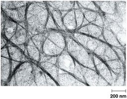
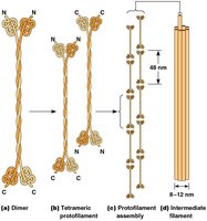
Intermediate filaments are organized into networks that confer mechanical strength and elasticity to tissues. They are dynamically remodeled in neurons and are less susceptible to chemical disruption than other cytoskeletal elements.
Tension-bearing: IFs withstand tensile forces.
Elasticity: IFs are flexible and resilient.
Integration of Cytoskeletal Elements
Mechanical Integration
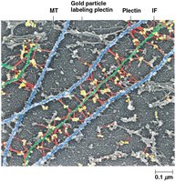
The cytoskeleton is a mechanically integrated structure, with microtubules resisting compression, microfilaments generating tension, and intermediate filaments providing elasticity. Proteins such as plectin connect these elements, ensuring coordinated cellular function.
Plectin: Links intermediate filaments to microfilaments and microtubules.
Integration: Essential for cellular stability and movement.