 Back
BackCytoskeletal Systems: Structure, Function, and Regulation
Study Guide - Smart Notes
 Tailored notes based on your materials, expanded with key definitions, examples, and context.
Cytoskeletal Systems
Overview of Cytoskeletal Elements
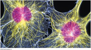
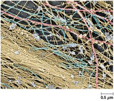
The cytoskeleton is a complex network of protein filaments that provides structural support, organization, and dynamic movement within eukaryotic cells. It consists of three primary types of elements: microtubules, microfilaments, and intermediate filaments, each with distinct properties and functions.
Microtubules: Rigid, hollow tubes composed of tubulin subunits (~25 nm diameter).
Microfilaments: Flexible, thin filaments composed of actin (~7 nm diameter).
Intermediate Filaments: Rope-like structures with variable composition (~8–12 nm diameter).
Septins: Additional polymer networks found in cells, composed of septin proteins.
Properties of Cytoskeletal Elements
Cytoskeletal elements are interconnected, highly structured, and dynamic. They are essential for cell shape, movement, and intracellular transport.
Rigid: Provides mechanical strength.
Highly-structured: Organized into elaborate networks.
Dynamic: Capable of rapid assembly and disassembly.
Interconnected: Elements interact with each other and with cellular structures.
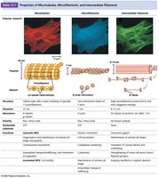
Comparison of Microtubules, Microfilaments, and Intermediate Filaments
The following table summarizes the main properties of the three cytoskeletal elements:
Property | Microtubules | Microfilaments | Intermediate Filaments |
|---|---|---|---|
Polymer Subunit | Tubulin | Actin | Varies (e.g., keratin, vimentin) |
Diameter | ~25 nm | ~7 nm | ~8–12 nm |
Structure | Hollow tube | Two-stranded helix | Rope-like |
Function | Cell shape, transport, mitosis | Muscle contraction, cell movement | Mechanical strength, structural support |
Microtubules
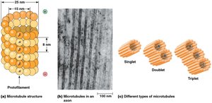
Structure and Types
Microtubules are straight, hollow cylinders composed of α- and β-tubulin heterodimers. They can form singlets, doublets, or triplets, depending on their cellular location and function.
Singlet Microtubules: Found in cytoplasm, consist of 13 protofilaments.
Doublet Microtubules: Found in cilia and flagella.
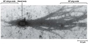
Triplet Microtubules: Found in basal bodies and centrioles.
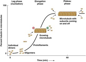
Assembly and Dynamics
Microtubule assembly occurs through nucleation and elongation phases, followed by a plateau phase where equilibrium is reached. The process is regulated by the concentration of tubulin and GTP.
Nucleation: Formation of oligomers that serve as seeds.
Elongation: Rapid addition of tubulin dimers.
Plateau: Structural equilibrium; assembly balanced by disassembly.
Critical Concentration: Tubulin concentration at which assembly equals disassembly.
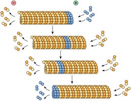
Polarity and Treadmilling
Microtubules have inherent polarity, with a plus end (fast-growing) and a minus end (slow-growing). Treadmilling occurs when subunit addition at the plus end and removal at the minus end are balanced.
Plus End: Rapid growth/shrinkage.
Minus End: Slow growth/shrinkage, often anchored at MTOC.
Treadmilling: Dynamic exchange of subunits.
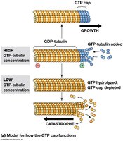
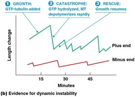
Dynamic Instability and GTP Hydrolysis
Microtubules exhibit dynamic instability, alternating between growth and shrinkage. GTP hydrolysis on β-tubulin regulates this process, with a GTP cap stabilizing the growing end.
GTP Cap: Prevents subunit loss; loss leads to catastrophe.
Catastrophe: Sudden transition from growth to shrinkage.
Rescue: Return to growth phase.
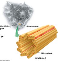
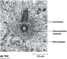
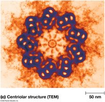
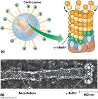
Microtubule-Organizing Centers (MTOCs) and Centrosomes
Microtubules originate from MTOCs, such as centrosomes, which contain centrioles and pericentriolar material. γ-tubulin ring complexes (γ-TuRCs) nucleate microtubule assembly.
Centrosome: Main MTOC in animal cells.
Centrioles: Nine triplet microtubules, involved in basal body formation.
γ-TuRCs: Nucleate new microtubules.
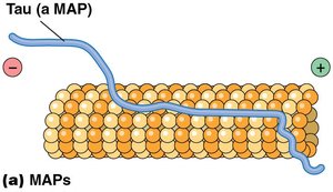
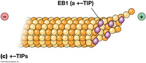
Microtubule-Binding Proteins
Microtubule stability and organization are regulated by various binding proteins:
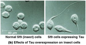
MAPs (Microtubule-Associated Proteins): Stabilize and bundle microtubules (e.g., Tau, MAP2).
+-TIP Proteins: Stabilize plus ends (e.g., EB1).
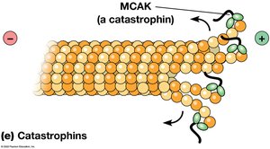
Catastrophins: Promote depolymerization (e.g., MCAK).
Microfilaments
Structure and Function
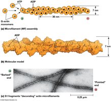
Microfilaments are the smallest cytoskeletal elements, composed of actin. They are crucial for muscle contraction, cell migration, cytokinesis, and cytoplasmic streaming.
Actin: Protein building block; exists as G-actin (globular) and F-actin (filamentous).
Polarity: Plus (barbed) end grows faster than minus (pointed) end.
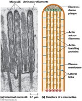
Cell Cortex and Microvilli
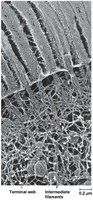
Microfilaments are concentrated beneath the plasma membrane at the cell cortex and form the structural core of microvilli, increasing surface area in intestinal cells.
Microvilli: Actin bundles crosslinked by proteins (fimbrin, villin).
Terminal Web: Network at base of microvillus, composed of myosin II and spectrin.
Actin-Binding Proteins and Regulation
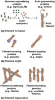
Actin polymerization, length, and organization are regulated by a variety of actin-binding proteins:
Monomer-sequestering proteins: Thymosin β4 binds G-actin, preventing polymerization.
Polymerizing proteins: Profilin promotes actin assembly.
Branching proteins: Arp2/3 complex nucleates actin branches, activated by WASP/WAVE.
Formins: Promote growth of long, unbranched actin filaments.
Capping proteins: CapZ (plus end), tropomodulin (minus end).
Severing proteins: Gelsolin breaks filaments and caps plus ends.
Crosslinking proteins: Filamin joins intersecting filaments.
Bundling proteins: α-actinin (focal adhesions), fascin (filopodia).
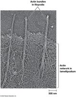
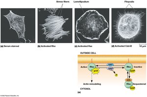
Actin Structures in Crawling Cells
Crawling cells exhibit specialized actin structures at their leading edge, including lamellipodia (branched actin network) and filopodia (parallel actin bundles).
Lamellipodia: Branched actin network for cell movement.
Filopodia: Parallel actin bundles for probing environment.
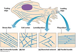

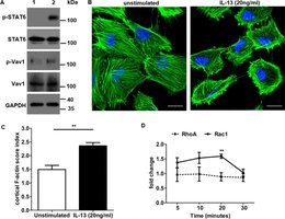
Regulation by Rho Family GTPases
Rho GTPases (Rho, Rac, Cdc42) regulate actin-based structures in response to cellular signals. They control formation of stress fibers, lamellipodia, and filopodia.
Rho: Promotes stress fiber formation.
Rac: Promotes lamellipodia extension.
Cdc42: Promotes filopodia formation.
Intermediate Filaments
Structure and Function
Intermediate filaments are the most stable and least soluble cytoskeletal components, providing mechanical strength and structural support. They are composed of fibrous proteins, such as keratin, vimentin, and lamins.
Keratin: Found in epithelial cells.
Vimentin: Found in mesenchymal cells.
Desmin: Found in muscle cells.
Lamins: Form nuclear scaffold.

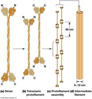
Assembly of Intermediate Filaments
IF proteins assemble from dimers into tetrameric protofilaments, which overlap to form a filamentous structure about eight protofilaments thick. The basic structural unit is a coiled-coil dimer.
Dimer: Two polypeptides intertwined.
Tetramer: Two dimers aligned laterally.
Filament: Eight protofilaments form the final structure.
Mechanical Integration of the Cytoskeleton
The cytoskeleton is a mechanically integrated structure, with microtubules resisting compression, microfilaments generating tension, and intermediate filaments withstanding tensile forces. Linker proteins such as spectraplakins (e.g., plectin) connect the three systems.
Spectraplakins: Link microtubules, microfilaments, and intermediate filaments.
Plectin: Found at sites of cytoskeletal interaction.

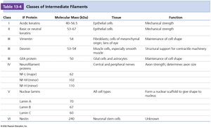
Summary Table: Classes of Intermediate Filaments
Class | IF Protein | Molecular Mass (kDa) | Tissue | Function |
|---|---|---|---|---|
I | Acidic keratins | 40–56.5 | Epithelial cells | Mechanical strength |
II | Basic/neutral keratins | 56–65 | Epithelial cells | Mechanical strength |
III | Vimentin, desmin, GFAP | 54 | Fibroblasts, muscle, glia | Cell shape, support |
IV | Neurofilament proteins | 68–240 | Neurons | Axon strength, diameter |
V | Lamins A, B, C | 70 | All cell types | Nuclear scaffold |
VI | Nestin | 240 | Neuronal stem cells | Unknown |
Conclusion
The cytoskeleton is essential for cell structure, function, and dynamics. Its three main components—microtubules, microfilaments, and intermediate filaments—work together to maintain cellular integrity, facilitate movement, and organize intracellular processes. Regulation by associated proteins and signaling pathways ensures precise control of cytoskeletal assembly and function.
