 Back
BackDNA, Chromosomes, and Sequencing: Cell Biology Study Guide
Study Guide - Smart Notes
 Tailored notes based on your materials, expanded with key definitions, examples, and context.
DNA: Structure and Function
Components of DNA
DNA (deoxyribonucleic acid) is the molecule that carries genetic information in cells. Its structure is fundamental to understanding genetics, heredity, and cellular function.
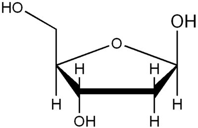
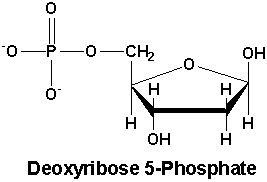
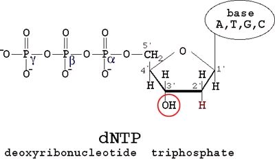
Sugar: The sugar in DNA is deoxyribose, a five-carbon sugar with carbons numbered 1' to 5'. The 'prime' notation distinguishes DNA carbons from those in the bases.
Phosphate Backbone: The phosphate group links the 5' carbon of one sugar to the 3' carbon of the next, forming the backbone of the DNA strand.
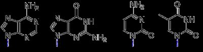
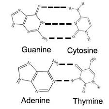
Bases: Four nitrogenous bases—adenine (A), guanine (G), cytosine (C), and thymine (T)—are attached to the sugar. Bases are classified as purines (A, G) and pyrimidines (C, T).
Nucleotides and DNA Strand Formation
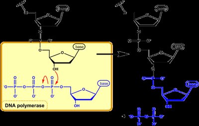
A nucleotide is the basic unit of DNA, consisting of a sugar, a phosphate, and a base. DNA polymerase catalyzes the formation of DNA strands by linking nucleotides in a 5' to 3' direction.
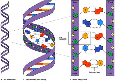
Directionality: DNA strands have direction, defined by the orientation of the sugar-phosphate backbone (5' to 3').
Polymerization: The 5' phosphate of a new nucleotide bonds to the 3' hydroxyl group of the existing strand.
Double-Stranded DNA and Base Pairing
DNA is typically double-stranded, with two complementary strands held together by hydrogen bonds between bases.
Base Pairing: Adenine pairs with thymine (A:T) via two hydrogen bonds; guanine pairs with cytosine (G:C) via three hydrogen bonds.
Strand Orientation: The two strands run in opposite directions (antiparallel).
Stability: G:C pairs are stronger due to three bonds, requiring more energy to separate.
Important Properties of DNA
Double-Stranded Preference: DNA prefers to be double-stranded; heat can denature it to single strands.
Information Storage: The sequence of bases encodes genetic information.
Mutations: Changes in base sequence (mutations) can lead to disease.
Accessing Information: Bases are on the inside of the helix, requiring cellular mechanisms to access genetic information.
Chromosomes: Structure and Analysis
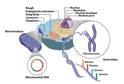
Nuclear vs. Mitochondrial DNA
Cells contain two types of DNA: nuclear DNA (in the nucleus) and mitochondrial DNA (in mitochondria).
Nuclear DNA: Highly compacted, organized into chromosomes, contains most genetic information.
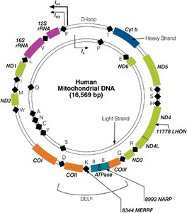
Mitochondrial DNA: Circular, double-stranded, encodes essential genes for mitochondrial function.

Chromosome Structure and Function
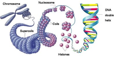
Chromosomes are DNA-protein complexes that package and protect genetic material.
Compaction: DNA is compacted ~10,000-fold to fit in the nucleus.
Protection: Chromosomes prevent DNA tangling and damage.
Replication and Division: Chromosomes ensure equal distribution of DNA during cell division.
Human Chromosome Number: Humans have 46 chromosomes in somatic cells (23 pairs).

Chromosome Appearance and Cell Cycle
Open State: Chromosomes are mostly open, allowing gene expression.
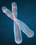
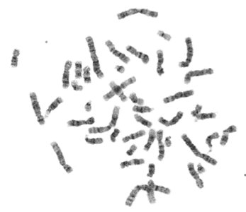
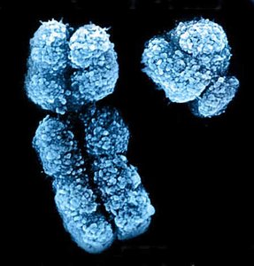
Metaphase State: Chromosomes are highly compacted and visible during metaphase.
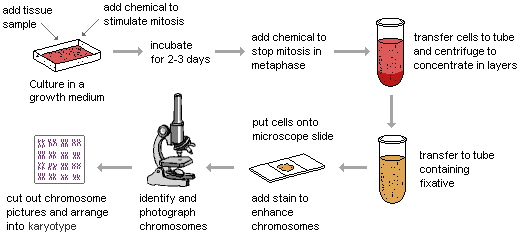
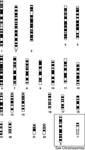
Chromosome Analysis: Karyotyping
Karyotyping is a laboratory technique to visualize and analyze chromosomes, especially during metaphase.
Metaphase Spread: Chromosomes are stained and photographed.
Karyotype: Chromosomes are arranged by size, banding pattern, and centromere position.
Applications: Detects large-scale genetic abnormalities (extra/missing material).
Chromosome Structure: Arms and Regions
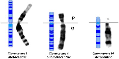
Centromere: Divides chromosome into short (p) and long (q) arms.
Telomeres: Protective ends of chromosomes.
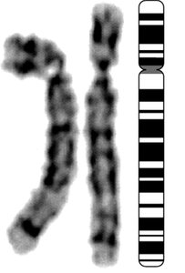
Banding: Staining reveals reproducible banding patterns for identification.
Chromosome Regions: Regions are named (e.g., 5q32) for precise location description.


Chromosome Classification by Centromere Position
Metacentric: Two arms of equal length.
Submetacentric: One short and one long arm.
Acrocentric: One visible arm; humans have five autosomal acrocentric chromosomes (13, 14, 15, 21, 22).
Human Chromosome Number and Types
Somatic Cells: 46 chromosomes (diploid: two sets).
Gametes: 23 chromosomes (haploid: one set).
Autosomes: 22 pairs (numbered by size).
Sex Chromosomes: X and Y; females have XX, males have XY.
Sex Chromosomes
X Chromosome: Large, gene-dense (~900 genes).
Y Chromosome: Small, few functional genes (~70), mostly related to male development.
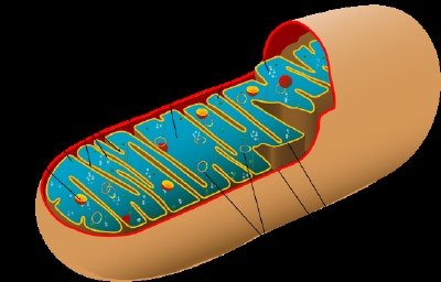
Mitochondrial DNA
Structure and Function
Mitochondria are the cell's energy factories, generating ATP via oxidative phosphorylation. Mitochondrial DNA is distinct from nuclear DNA.
Genome: Circular, double-stranded, 16,500 kb, 37 genes (2 rRNAs, 22 tRNAs, 13 polypeptides).
Inheritance: Mitochondrial DNA is maternally inherited.
Independence: Mitochondria replicate independently and do not appear in karyotype analysis.
Sanger Sequencing and DNA Analysis
Principles of Sanger Sequencing
Sanger sequencing is a method to determine the order of bases in a DNA template, based on PCR with modifications.
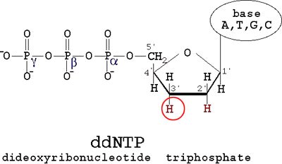
Key Steps: Uses one primer, incorporates dideoxynucleotides (ddNTPs) with colored fluorophores.
Chain Termination: ddNTPs lack a 3' hydroxyl, causing chain termination when incorporated.
Sequence Determination: The resulting fragments are separated and detected by color, revealing the DNA sequence.
Applications and Mutation Detection
Mutation Analysis: Sanger sequencing can detect single nucleotide changes, insertions, and deletions.
Limitations: Does not indicate the chromosomal origin of the PCR product.
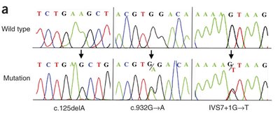
Table: Comparison of Nuclear and Mitochondrial DNA
Feature | Nuclear DNA | Mitochondrial DNA |
|---|---|---|
Location | Nucleus | Mitochondria |
Structure | Linear, multiple chromosomes | Circular, single molecule |
Size | ~3 billion base pairs | ~16,500 base pairs |
Gene Content | ~20,000-25,000 genes | 37 genes |
Inheritance | Biparental | Maternally inherited |
Compaction | Highly compacted (chromosomes) | Not compacted at metaphase |
Appearance in Karyotype | Yes | No |
Table: DNA Base Pairing
Base 1 | Base 2 | Bond Type | Number of Bonds |
|---|---|---|---|
Adenine (A) | Thymine (T) | Hydrogen | 2 |
Guanine (G) | Cytosine (C) | Hydrogen | 3 |
Table: Chromosome Types by Centromere Position
Type | Description | Example |
|---|---|---|
Metacentric | Arms equal length | Chromosome 1 |
Submetacentric | One short, one long arm | Chromosome 4 |
Acrocentric | One visible arm | Chromosome 14 |
Summary
This guide covers the structure and function of DNA, chromosomes, and mitochondrial DNA, as well as the principles and applications of Sanger sequencing. Understanding these concepts is essential for cell biology, genetics, and molecular biology studies.
