 Back
BackElectron Transport Chain and ATP Synthesis in Mitochondria
Study Guide - Smart Notes
 Tailored notes based on your materials, expanded with key definitions, examples, and context.
Mitochondrial Structure and Function
Overview of Mitochondria
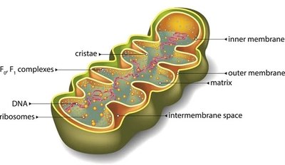
Mitochondria are dynamic, double-membraned organelles found in nearly all eukaryotic cells. They are the primary site of ATP production through aerobic respiration and play a central role in cellular energy metabolism.
Double membrane system: Consists of an outer membrane (with porins for permeability) and a highly folded inner membrane (cristae) that increases surface area for metabolic reactions.
Matrix: The innermost compartment containing enzymes for the TCA cycle, mitochondrial DNA, and ribosomes.
Endosymbiotic hypothesis: Suggests mitochondria originated from ancestral prokaryotes engulfed by early eukaryotic cells.
Mitochondrial Membranes and Compartments
The inner mitochondrial membrane is less permeable and contains the protein complexes of the electron transport chain (ETC) and ATP synthase. The intermembrane space is the region between the inner and outer membranes, crucial for proton gradient formation.
Cristae: Infoldings of the inner membrane that house ETC components and ATP synthase, maximizing surface area for energy conversion.
Matrix: Contains TCA cycle enzymes, mitochondrial DNA, and ribosomes, supporting semi-autonomous function.
Electron Transport Chain (ETC)
Overview of the ETC
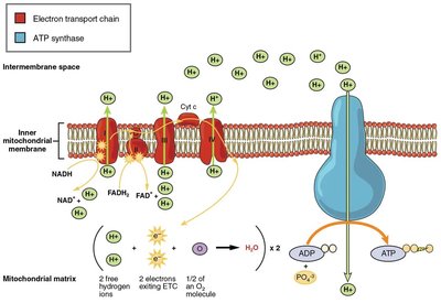
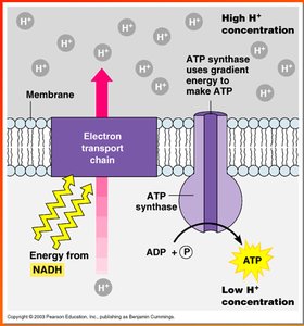
The electron transport chain is a series of protein complexes and electron carriers embedded in the inner mitochondrial membrane. It transfers electrons from NADH and FADH2 to molecular oxygen, generating a proton gradient used for ATP synthesis.
Electron donors: NADH and FADH2 generated from glycolysis and the TCA cycle.
Final electron acceptor: Molecular oxygen (O2), which is reduced to water.
Proton gradient: Electron transfer is coupled to the active transport of H+ ions from the matrix to the intermembrane space, creating an electrochemical gradient.
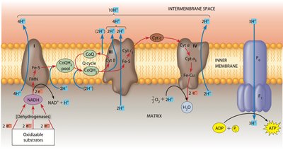
Major Components of the ETC
Complex I (NADH: ubiquinone oxidoreductase): Receives electrons from NADH, passes them to coenzyme Q (CoQ), and pumps 4 H+ into the intermembrane space.
Complex II (Succinate dehydrogenase): Receives electrons from FADH2 (via succinate), passes them to CoQ, but does not pump protons.
Complex III (Cytochrome bc1 complex): Transfers electrons from CoQH2 to cytochrome c and pumps protons.
Complex IV (Cytochrome c oxidase): Transfers electrons from cytochrome c to O2, forming water and pumping protons.
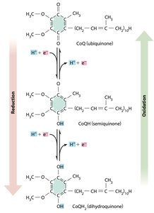
Mobile carriers: Coenzyme Q (ubiquinone) and cytochrome c shuttle electrons between complexes.
Electron Carriers in the ETC
Electron carriers facilitate the stepwise transfer of electrons, maximizing energy capture and minimizing heat loss.
Flavoproteins: Proteins with flavin groups (FMN or FAD) that transfer electrons and protons.
Iron-sulfur (Fe-S) proteins: Transfer electrons only, coordinated by cysteine residues.
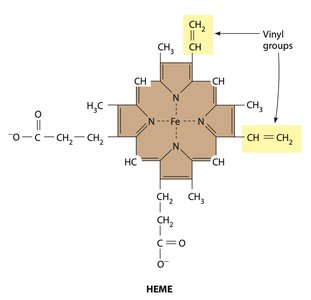
Cytochromes: Proteins with heme groups (Fe center) that transfer electrons; some contain copper (Cu) centers for O2 reduction.
Coenzyme Q (ubiquinone): A lipid-soluble, non-protein carrier that accepts and donates electrons and protons.
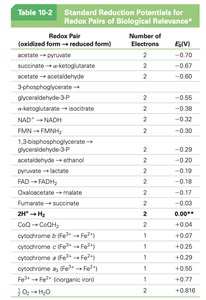
Redox Potential and Free Energy
Electron flow through the ETC is driven by differences in redox potential (E0'), with electrons moving from carriers with lower (more negative) to higher (more positive) potentials. This flow is coupled to free energy changes (ΔG) that drive proton pumping.
Redox potential (E0'): Indicates a molecule's tendency to accept electrons; higher values mean stronger oxidizing agents.
Free energy change: Calculated by the Nernst equation:
Where n = number of electrons transferred, F = Faraday constant (23.2 kcal/V·mol), and ΔE = difference in redox potential.
ATP Synthesis: Chemiosmosis and ATP Synthase
Chemiosmotic Theory
Chemiosmosis describes the process by which the energy stored in a proton gradient across the inner mitochondrial membrane is used to drive ATP synthesis. The ETC establishes this gradient by pumping protons into the intermembrane space.
Proton-motive force: The electrochemical gradient of H+ ions provides the energy for ATP synthesis.
ATP yield: Approximately 3 ATP per NADH and 2 ATP per FADH2 oxidized.
ATP Synthase Structure and Function
ATP synthase (F1F0 complex) is a multi-subunit enzyme embedded in the inner mitochondrial membrane. It synthesizes ATP from ADP and inorganic phosphate (Pi) as protons flow back into the matrix through its channel.
F0 subunit: Forms a proton channel across the membrane.
F1 subunit: Contains catalytic sites for ATP synthesis, protruding into the matrix.
Rotational catalysis: Proton flow drives rotation of the γ subunit, inducing conformational changes in the β subunits to catalyze ATP formation (binding change model).

Binding Change Model of ATP Synthesis
The binding change model explains how ATP synthase produces ATP. As protons move through the F0 channel, the γ subunit rotates, causing the β subunits of F1 to cycle through three conformations: binding ADP and Pi, catalyzing ATP synthesis, and releasing ATP.
Three β subunits: Each in a different state (open, loose, tight) at any time.
Rotation: Driven by proton flow, essential for ATP production.
Regulation and Inhibition of the ETC
Uncoupling Agents
Certain chemicals, such as 2,4-dinitrophenol (DNP), uncouple electron transport from ATP synthesis by dissipating the proton gradient. This results in energy being released as heat instead of being used for ATP production, which can be fatal.
DNP: Was used as a weight loss drug but caused dangerous overheating and death due to uncontrolled proton flow and heat generation.
Clinical relevance: Uncouplers highlight the importance of the proton gradient in cellular energy metabolism.
Summary Table: Electron Transport Chain Complexes
Complex | Electron Source | Electron Carrier | Protons Pumped | Final Electron Acceptor |
|---|---|---|---|---|
I | NADH | FMN, Fe-S, CoQ | 4 | CoQ |
II | FADH2 (succinate) | FAD, Fe-S, CoQ | 0 | CoQ |
III | CoQH2 | Cytochrome b, Fe-S, cytochrome c | 4 | Cytochrome c |
IV | Cytochrome c | Cytochrome a, a3, Cu | 2 | O2 (forms H2O) |
Key Equations
Free energy change for electron transfer:
ATP yield per electron carrier:
1 NADH ≈ 3 ATP 1 FADH2 ≈ 2 ATP
Additional info:
Some images and tables were inferred to clarify the structure and function of the ETC and ATP synthase. The summary table was constructed based on standard cell biology knowledge of the ETC complexes.
