 Back
BackGene Expression and the Genetic Code: From Transcription to Translation and Mutation
Study Guide - Smart Notes
 Tailored notes based on your materials, expanded with key definitions, examples, and context.
HIV Life Cycle and Host Interaction
Viral Entry and Reverse Transcription
The HIV life cycle begins with the entry of the virus into the host cell, followed by the reverse transcription of its RNA genome into DNA. This process is catalyzed by the viral enzyme reverse transcriptase.
Viral Entry: HIV binds to specific receptors and co-receptors on the host cell membrane, facilitating fusion and entry.
Reverse Transcription: The viral RNA is reverse transcribed into complementary DNA (cDNA), which is then integrated into the host genome.
Integration: The newly synthesized viral DNA is transported into the nucleus and integrated into the host's chromosomal DNA by the viral integrase enzyme.
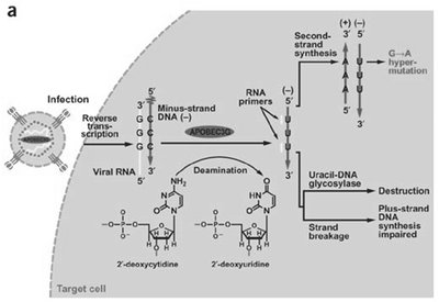
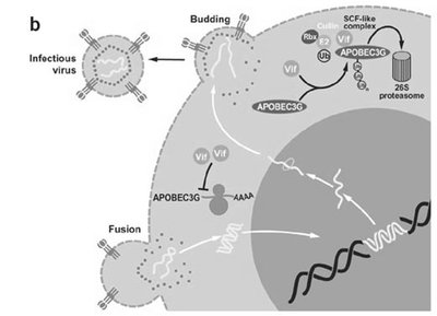
Host Defense: APOBEC3G and Viral Counteraction
APOBEC3G is a host cytidine deaminase that can edit the HIV genome, introducing mutations that impair viral replication. However, the viral protein Vif counteracts APOBEC3G by targeting it for degradation.
APOBEC3G Function: Deaminates cytidine to uridine in viral DNA, leading to G-to-A hypermutations and defective viruses.
Vif Protein: HIV Vif binds APOBEC3G and recruits an E3 ubiquitin ligase complex, leading to APOBEC3G degradation via the proteasome.
Gene Expression: From DNA to Protein
Transcription and RNA Processing
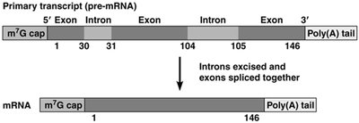
Gene expression begins with the transcription of DNA into pre-mRNA, which contains both exons (coding regions) and introns (non-coding regions). The pre-mRNA undergoes processing to become mature mRNA.
5' Capping: Addition of a 7-methylguanosine cap to the 5' end of the pre-mRNA.
Splicing: Removal of introns and joining of exons to form a continuous coding sequence.
Polyadenylation: Addition of a poly(A) tail at the 3' end for stability and export.
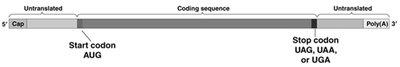
mRNA Structure and Open Reading Frames (ORFs)
The mature mRNA contains untranslated regions (UTRs) at both ends, a coding sequence that begins with a start codon (AUG), and ends with a stop codon (UAG, UAA, or UGA).
Start Codon (AUG): Specifies the initiation of translation and encodes methionine.
Stop Codons: Signal the termination of translation.
ORF: The open reading frame is the stretch of codons between the start and stop codons.
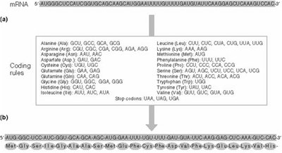
The Genetic Code
The genetic code is a set of rules by which the nucleotide sequence of mRNA is translated into the amino acid sequence of proteins. It is a triplet code, meaning each amino acid is encoded by a sequence of three nucleotides (codon).
Degeneracy: Most amino acids are encoded by more than one codon.
Nonoverlapping: Each nucleotide is part of only one codon.
Universal: The genetic code is nearly universal among organisms, with minor variations.
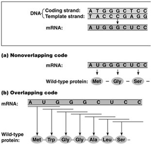
Nonoverlapping vs. Overlapping Codes
In the standard genetic code, codons are read in a nonoverlapping manner. Overlapping codes would allow a single nucleotide to be part of multiple codons, but this is not the case in most organisms.
Nonoverlapping: Each codon is read sequentially, three nucleotides at a time.
Overlapping (hypothetical): Each nucleotide could be part of more than one codon, leading to different reading frames.
Frameshift Mutations
Frameshift mutations occur when nucleotides are inserted or deleted from the DNA sequence in numbers not divisible by three, altering the reading frame and potentially changing every amino acid downstream of the mutation.
Effect: Can result in nonfunctional proteins due to extensive missense or premature stop codons.

Mutations and Their Effects on Proteins
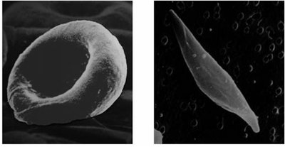
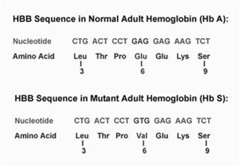
Sickle Cell Anemia: A Case Study
Sickle cell anemia is caused by a single nucleotide mutation in the β-hemoglobin gene (HBB), resulting in the substitution of valine for glutamic acid in the protein. This leads to abnormal hemoglobin structure and sickle-shaped red blood cells.
Mutation: A to T transversion in DNA, resulting in Glu to Val substitution in the protein.
Consequence: Hemoglobin molecules aggregate, distorting red blood cell shape and causing anemia.
Types of Mutations
Silent Mutation: A base substitution that does not change the encoded amino acid due to the degeneracy of the genetic code.
Missense Mutation: A base substitution that changes one amino acid to another.
Nonsense Mutation: A base substitution that introduces a premature stop codon, leading to truncated proteins.
Frameshift Mutation: Insertion or deletion of nucleotides not in multiples of three, altering the reading frame.
Translation: From mRNA to Protein
Translation Machinery
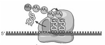
Translation is the process by which ribosomes synthesize proteins using mRNA as a template. The main components are mRNA, ribosomes, tRNA, aminoacyl-tRNA synthetases, and translation factors.
mRNA: Provides the template for the amino acid sequence.
Ribosome: The molecular machine that catalyzes peptide bond formation.
tRNA: Adaptor molecules that bring amino acids to the ribosome and recognize codons via their anticodon loop.
Aminoacyl-tRNA Synthetase: Enzymes that attach the correct amino acid to its corresponding tRNA.
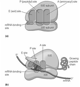
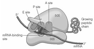
Ribosome Structure and Function
Ribosomes have three important binding sites for tRNA: the A (aminoacyl), P (peptidyl), and E (exit) sites. Translation proceeds in the 5' to 3' direction along the mRNA, synthesizing the polypeptide from the N-terminus to the C-terminus.
A Site: Entry point for aminoacyl-tRNA.
P Site: Holds the tRNA with the growing peptide chain.
E Site: Exit site for deacylated tRNA.
The Wobble Hypothesis
The wobble hypothesis explains how a single tRNA can recognize multiple codons for the same amino acid, due to flexible base pairing at the third codon position.
Inosine: A modified base in tRNA that can pair with U, C, or A in the mRNA codon.
Result: Fewer than 61 tRNAs are needed to recognize all sense codons.

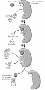
Aminoacyl-tRNA Synthetase and tRNA Charging
Aminoacyl-tRNA synthetases are enzymes that attach the correct amino acid to its corresponding tRNA, a process known as "charging." This reaction is ATP-dependent and forms a high-energy ester bond.
Specificity: Each aminoacyl-tRNA synthetase is specific for one amino acid and its compatible tRNAs.
Charged tRNA: The product is an aminoacyl-tRNA, ready for translation.
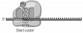
Translation Initiation, Elongation, and Termination
Translation occurs in three main stages: initiation, elongation, and termination.
Initiation: The small ribosomal subunit binds to the mRNA and the initiator tRNA (carrying methionine) pairs with the start codon. In eukaryotes, this process involves scanning for the Kozak sequence.
Elongation: Amino acids are sequentially added to the growing polypeptide chain as the ribosome moves along the mRNA.
Termination: When a stop codon is encountered, release factors promote the release of the completed polypeptide and dissociation of the ribosome.
Protein Folding and Intracellular Sorting
Protein Folding and Chaperones
Newly synthesized polypeptides begin folding during translation. Molecular chaperones, such as Hsp70 and Hsp60, assist in proper folding and prevent aggregation.
Chaperonins: Specialized proteins that provide an isolated environment for folding.
Misfolding: Can lead to diseases such as Alzheimer's and mad cow disease.
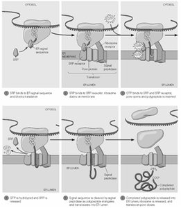
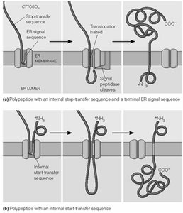
Protein Targeting and Cotranslational Import
Proteins destined for secretion or for certain organelles contain signal peptides that direct their cotranslational import into the endoplasmic reticulum (ER). The signal recognition particle (SRP) recognizes the signal sequence and facilitates targeting to the ER membrane.
Signal Peptide: Short, hydrophobic sequence at the N-terminus of the polypeptide.
SRP: Binds the signal peptide and pauses translation until the ribosome docks at the ER.
Cleavage: The signal peptide is removed after import into the ER lumen.
Summary Table: Types of Mutations and Their Effects
Mutation Type | Definition | Effect on Protein |
|---|---|---|
Silent | Base substitution with no amino acid change | No effect |
Missense | Base substitution changes one amino acid | May alter protein function |
Nonsense | Base substitution introduces stop codon | Truncated, usually nonfunctional protein |
Frameshift | Insertion/deletion not in multiples of three | Extensive missense, premature stop codon |
