 Back
BackHuman Genetics: Mendelian and Non-Mendelian Inheritance, Chromosomal Theory, and Genetic Diseases
Study Guide - Smart Notes
 Tailored notes based on your materials, expanded with key definitions, examples, and context.
Genetics and Eye Color
Heterochromia and Melanin Distribution
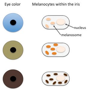
Eye color is determined by the amount and distribution of melanin in the iris, produced by melanocytes. Heterochromia occurs when each iris has a different amount of melanin, resulting in distinct eye colors. Melanin is produced by melanocytes, and pre-melanocytes must migrate to the eyes during development. Damage to melanocytes leads to reduced melanin and lighter eye color.
Most melanin: dark brown eyes
Least melanin: blue eyes
Hazel and green eyes: intermediate melanin levels
Genetic variations affect melanin production and distribution
Genetic Basis of Eye Color
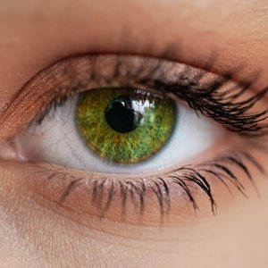
Eye color is a polygenic trait, meaning it is controlled by multiple genes. For example, green eyes are rare globally but common in certain populations, and at least 16 genes have been identified as contributing to eye color.
Hazel eyes: more melanin than green, less than brown
Green eyes: medium melanin, 2% worldwide, 86% in Ireland/Scotland/Northern Europe
Polygenic inheritance leads to a wide variety of eye colors

Chromosomal Theory of Inheritance
Basic Principles
The chromosomal theory of inheritance states that nuclei of all somatic cells contain two sets of homologous chromosomes, one maternal and one paternal. Chromosomes retain their individuality throughout the organism's life cycle, and homologous chromosomes are functionally equivalent except for their alleles.
Maternal and paternal homologs synapse during meiosis and segregate into different cells
Homologous pairs segregate independently during meiosis
Mendel’s Laws and Their Limitations
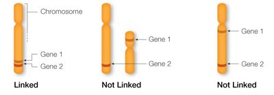
Mendel’s laws, such as the law of independent assortment, apply only when genes are on separate chromosomes and are not linked. Many traits are controlled by multiple genes, linked genes, or genes on sex chromosomes, which can violate Mendel’s laws.
Linked genes: inherited together, not sorted independently
Polygenic traits: controlled by multiple genes
Sex-linked traits: located on X or Y chromosomes

Mendelian Genetics: Dihybrid Crosses
Law of Independent Assortment
The law of independent assortment states that the alleles of different genes segregate independently during gamete formation. This results in all possible combinations of traits appearing in offspring.
Example: Dihybrid cross between Ttpp (tall, white) and ttPp (short, purple)
Genotypic ratio: 1:1:1:1 (TtPp, ttPp, Ttpp, ttpp)
Phenotypic ratio: 1:1:1:1 (tall & purple, short & purple, tall & white, short & white)
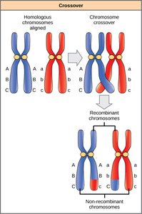
Linked Genes and Recombination
Linked genes are located close together on the same chromosome and are usually inherited together. Crossing over during meiosis can disrupt linkage, and the frequency of recombination depends on the distance between genes.
Linked genes violate the law of independent assortment
Recombination frequency increases with gene distance
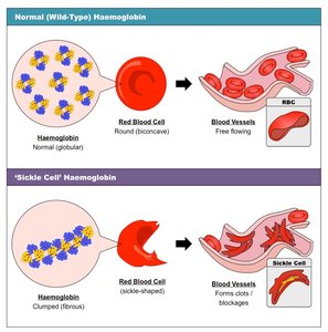
Genetic Diseases: Sickle Cell Anemia
Case Study: Sickle Cell Anemia
Sickle cell anemia is caused by a single amino acid mutation in the gene encoding the β subunit of hemoglobin. This mutation changes the shape of red blood cells from round (globular) to sickle-shaped (fibrous), leading to anemia, poor blood flow, and reduced oxygen transport.
Normal β-globin: βA
Sickle β-globin: βS
Autosomal recessive disease: requires two βS alleles
Heterozygous (βAβS): sickle cell trait, provides evolutionary advantage against malaria

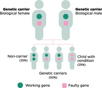
Inheritance Patterns
Both parents can be carriers (heterozygous βAβS), and the chance of offspring having sickle cell disease follows Mendelian ratios.
Genotypic ratio: 1 βAβA : 2 βAβS : 1 βSβS
Phenotypic ratio: 3 unaffected/trait : 1 affected

Carrier Screening
Carrier screening is used to test for autosomal and X-linked recessive diseases before conception. Examples include cystic fibrosis, PKU, and Tay-Sachs disease.
Genetic Diseases: Hemophilia
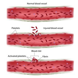
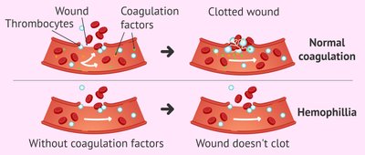
Hemophilia and Blood Clotting
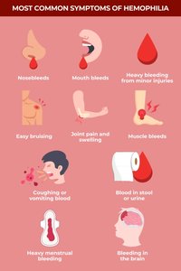
Hemophilia is an inherited blood clotting disorder caused by mutations in genes encoding clotting factor proteins. These proteins work with platelets to form clots and stop bleeding. Hemophilia can be mild, moderate, or severe, with symptoms ranging from prolonged bleeding to spontaneous internal bleeding.
Hemophilia A: F8 gene (factor VIII)
Hemophilia B: F9 gene (factor IX)
Hemophilia C: rare
X-linked Inheritance
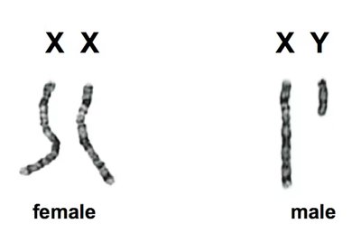
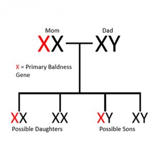
Hemophilia is an X-linked recessive disease. Biological males (XY) have only one X chromosome, so a single mutated allele causes the disease. Biological females (XX) require two mutated alleles to be affected, but carriers can sometimes show symptoms.
X-linked traits: red-green color blindness, male-pattern baldness, Duchenne muscular dystrophy
Y-linked traits: only inherited from XY parent, no dominant/recessive pattern
Webbed Toes and Y-linked Traits
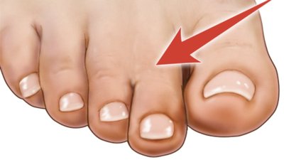
Webbed Toes (Syndactyly)
Webbed toes are a type of syndactyly, caused by incomplete apoptosis during development. It can be inherited as a Mendelian trait on an autosome or as a sex-linked trait on the Y chromosome.
Occurs in ~1/2,500 live births
Y-linked traits: only present in people with Y chromosome
Y chromosome: contains ~150 genes, including SRY gene for sex determination

Sex Chromosomes and Biological Sex
Sex Determination
Eggs always contribute an X chromosome, while sperm can contribute either X or Y. The SRY gene on the Y chromosome is the master switch for male-typical sexual development. Extra, missing, or rearranged sex chromosomes can lead to syndromes such as Turner (X), Klinefelter (XXY), and XYY syndrome.
Intersex: internal/external sex characteristics outside traditional male/female binaries
All human fetuses begin with undifferentiated genitalia
SRY gene activates around 9 weeks gestation
Non-Mendelian Genetics: Incomplete Dominance and Co-dominance
Case Study: Snapdragon Flowers (Antirrhinum majus)
Snapdragons demonstrate incomplete dominance, where both alleles are partially expressed, resulting in an intermediate phenotype. The F1 generation is a hybrid, and the F2 generation shows a mix of parental and intermediate phenotypes.
Genotypic ratio: 1:2:1 (RR, Rr, rr)
Phenotypic ratio: 1 red : 2 pink : 1 white
Case Study: ABO Blood Types
ABO blood types are determined by the structure of carbohydrates on red blood cell membranes. The ABO gene encodes a glycosyltransferase enzyme, and the alleles IA and IB are co-dominant, meaning both are expressed in heterozygotes. O blood type is recessive (ii).
A: N-acetylgalactosamine
B: galactose
AB: both sugars present
O: no glycosylation
Polygenic Traits: Labrador Retriever Coat Color
Polygenic Inheritance
Labrador retriever coat color is controlled by multiple genes (polygenic). MC1R determines yellow color, and TYRP1 determines black or chocolate color. Black is dominant over chocolate, and the effect of TYRP1 depends on MC1R.
MC1R: E = black/chocolate, e = yellow
TYRP1: B = black, b = chocolate
Yellow labs (ee) only have yellow puppies
Black/chocolate labs can produce yellow puppies if heterozygous for MC1R
Polygenic traits often show partial phenotypes, co-dominance, and incomplete dominance, making non-Mendelian genetics common in nature.
