 Back
Back19: Intracellular Transport and Protein Sorting in Eukaryotic Cells
Study Guide - Smart Notes
 Tailored notes based on your materials, expanded with key definitions, examples, and context.
Protein Sorting and Intracellular Membrane Trafficking
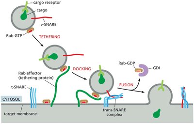
Vesicle Tethering, Docking, and Fusion
Intracellular transport relies on the precise delivery of vesicles to their target membranes, a process orchestrated by specific protein families. The steps include tethering, docking, and fusion, ensuring that cargo is delivered to the correct cellular compartment.
Tethering: Rab proteins (monomeric GTPases) and Rab effectors guide vesicles to their target membranes.
Docking: SNARE proteins (v-SNARE on vesicle, t-SNARE on target) interact to bring membranes close together.
Fusion: SNARE regulators mediate the fusion of lipid bilayers, allowing cargo delivery. Rab-GDP dissociation inhibitor (GDI) recycles Rab proteins.
Example: Vesicle carrying cargo fuses with the plasma membrane to release its contents.
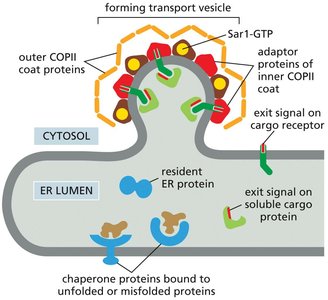
Recruitment of Cargo into ER Transport Vesicles
Proteins destined for secretion or for other organelles exit the endoplasmic reticulum (ER) in COPII-coated vesicles. The selection of cargo is highly regulated to ensure only properly folded proteins are transported.
Cargo Selection: Adaptor proteins recognize exit signals on cargo receptors and soluble cargo proteins.
Coat Proteins: Sar1-GTP initiates the assembly of the COPII coat, which shapes the vesicle.
Quality Control: Chaperone proteins retain unfolded or misfolded proteins in the ER.
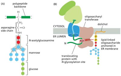
Protein Modification and Processing in the ER and Golgi Apparatus
N-linked Glycosylation in the Rough ER
Most proteins entering the ER are covalently modified by the addition of oligosaccharides, a process known as N-linked glycosylation. This modification is critical for protein folding, stability, and sorting.
Attachment Site: The precursor oligosaccharide is attached to asparagine (Asn) residues in the sequence Asn-X-Ser or Asn-X-Thr.
Enzyme: Oligosaccharyl transferase, a membrane-bound enzyme, catalyzes this reaction.
Further Processing: Extensive trimming and modification occur in the Golgi apparatus.
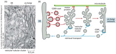
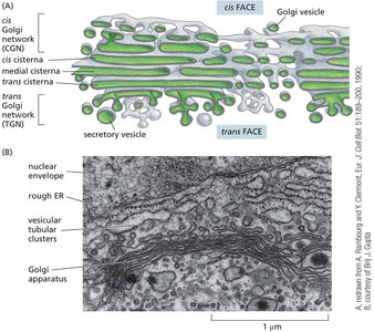
Vesicular Transport from ER to Golgi Apparatus
Proteins are transported from the ER to the Golgi apparatus via vesicular tubular clusters. These clusters move along microtubules and facilitate both forward and retrieval transport.
Forward Transport: COPII-coated vesicles carry proteins from the ER to the Golgi.
Retrieval Transport: COPI-coated vesicles return escaped ER proteins and vesicle components back to the ER.
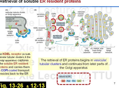
Retrieval of Soluble ER Resident Proteins
Some proteins that escape the ER are retrieved from the Golgi and returned to the ER. This retrieval is mediated by the KDEL receptor, which recognizes a specific amino acid sequence (KDEL) on ER-resident proteins.
KDEL Sequence: Lys-Asp-Glu-Leu at the C-terminus signals return to the ER.
Transport: COPI-coated vesicles carry these proteins back to the ER.
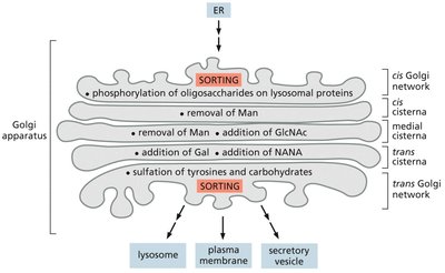
Structure and Function of the Golgi Apparatus
The Golgi apparatus is a series of flattened membrane-bound compartments where proteins undergo further modification, sorting, and packaging for delivery to their final destinations.
Organization: The Golgi has cis (entry) and trans (exit) faces, with medial cisternae in between.
Protein Fate: Proteins are modified and sorted as they move from the cis to the trans face.
Oligosaccharide Processing in Golgi Compartments
As proteins traverse the Golgi, their oligosaccharide chains are sequentially modified by specific enzymes localized to different cisternae.
Early-acting Enzymes: Located in the cis Golgi cisternae (e.g., removal of mannose).
Late-acting Enzymes: Located in the trans Golgi cisternae (e.g., addition of galactose, sialic acid).
Sorting: Proteins are sorted for delivery to lysosomes, the plasma membrane, or secretory vesicles.
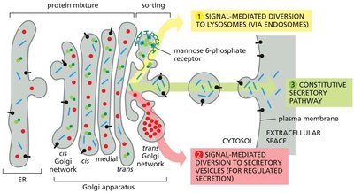
Protein Sorting in the Trans Golgi Network
The trans Golgi network (TGN) is the major sorting station where proteins are directed into one of several pathways based on their final destination.
Constitutive Pathway: Operates in all cells, continuously delivering proteins and lipids to the plasma membrane.
Regulated Pathway: Used by specialized cells to store secretory proteins in vesicles for release upon stimulation.
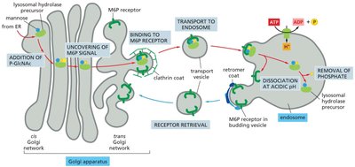
Lysosomal Pathway: Delivers enzymes to lysosomes via mannose-6-phosphate tagging.
Transport to Lysosomes and Secretory Vesicle Formation
Transport of Lysosomal Hydrolases
Lysosomal enzymes are tagged with mannose-6-phosphate (M6P) in the Golgi, which directs them to endosomes and ultimately lysosomes.
M6P Tag: Added in the cis Golgi, recognized by M6P receptors in the TGN.
Delivery: Enzymes are transported in clathrin-coated vesicles to endosomes, where the acidic environment releases the enzyme.
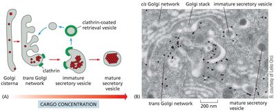
Formation and Exocytosis of Secretory Vesicles
Secretory vesicles form from the TGN, concentrating their protein cargo. Upon receiving a signal, these vesicles fuse with the plasma membrane to release their contents (exocytosis).
Cargo Concentration: Secretory proteins are highly concentrated in vesicles.
Stimulus-Dependent Release: Fusion with the plasma membrane is triggered by specific signals, such as increased intracellular Ca2+.
Example: Insulin release from pancreatic β cells.
Endocytic Pathways
Types of Endocytosis
Endocytosis is the process by which cells internalize substances from their environment. There are several types, each with distinct mechanisms and functions.
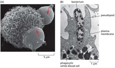
Phagocytosis: Ingestion of large particles (e.g., microorganisms, cell debris) via large vesicles called phagosomes. Important for immune defense and tissue homeostasis.
Pinocytosis: Uptake of fluids and small molecules via small vesicles.
Receptor-Mediated Endocytosis: Selective uptake of specific macromolecules bound to cell surface receptors, often via clathrin-coated vesicles (e.g., LDL, vitamin B12, iron).
Endosome Maturation and Lysosomal Degradation
Endocytosed material is delivered to endosomes, which mature from early to late forms and eventually fuse with lysosomes for degradation.
Early Endosomes: Located near the plasma membrane, act as sorting stations.
Late Endosomes: Move toward the cell center, lose tubular projections, and prepare for fusion with lysosomes.
Lysosomes: Degrade macromolecules delivered via multiple pathways, including endocytosis, phagocytosis, and autophagy.
Summary Table: Major Vesicular Transport Pathways
Pathway | Coat Protein | Direction | Main Cargo |
|---|---|---|---|
ER to Golgi | COPII | Forward (anterograde) | Secretory and membrane proteins |
Golgi to ER | COPI | Retrieval (retrograde) | ER-resident proteins, vesicle components |
TGN to Lysosome | Clathrin | Sorting | Lysosomal hydrolases |
TGN to Plasma Membrane | None (constitutive/regulated) | Secretion | Secretory proteins, membrane proteins |
Additional info: This guide covers key aspects of intracellular transport, protein sorting, and endocytic pathways, integrating foundational cell biology concepts with visual aids for enhanced understanding.
