 Back
BackIntroduction to Cell and Molecular Biology: Cell Theory, Microscopy, and Cell Types
Study Guide - Smart Notes
 Tailored notes based on your materials, expanded with key definitions, examples, and context.
Cell Theory
Foundations of Cell Theory
The cell theory is a fundamental concept in biology that describes the properties of cells, the basic unit of life. It was developed in the 19th century and remains central to our understanding of living organisms.
All organisms consist of one or more cells (Schwann, 1839): Every living thing is made up of cells, whether unicellular or multicellular.
The cell is the basic unit of structure for all organisms (Schwann, 1839): Cells are the smallest units that can carry out all life processes.
All cells arise from preexisting cells (Virchow, 1855): Cells do not spontaneously appear; they are produced by the division of existing cells.
Additional info: The cell theory laid the groundwork for modern cell biology, emphasizing the continuity of life and the importance of cellular organization.
Microscopy and the Study of Cells
Resolution and Limitations of Light Microscopy
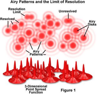
Microscopy is essential for studying cells, as most are too small to be seen with the naked eye. The resolution of a microscope is the shortest distance between two points that can still be distinguished as separate entities.
Resolution of light microscopes: About 0.2 microns (μm), limited by the wavelength of visible light ().
Limitations: Only dark objects are seen well, out-of-focus light reduces clarity, and the maximum useful magnification is about 1000x.
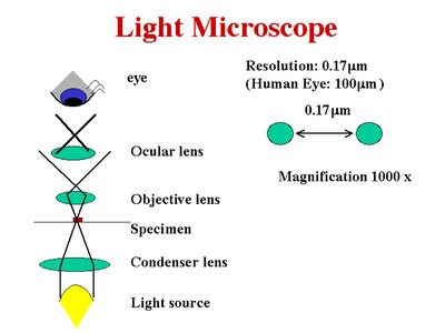
Additional info: The human eye can resolve objects down to about 100 μm, while light microscopes extend this to about 0.2 μm.
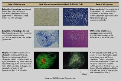
Types of Microscopy
Different microscopy techniques allow visualization of various cellular structures and molecules.
Type of Microscopy | Description | Application |
|---|---|---|
Brightfield (unstained) | Light passes directly through specimen; little contrast unless naturally pigmented or artificially stained. | General cell observation |
Brightfield (stained) | Staining increases contrast; cells must be fixed (preserved). | Detailed cell structure |
Phase contrast | Enhances contrast in transparent specimens without staining. | Live cell imaging |
Differential interference contrast | Uses optical modifications to exaggerate differences in refractive index. | 3D-like images of cells |
Fluorescence | Shows locations of specific molecules using fluorescent dyes or proteins. | Localization of proteins, organelles |
Confocal | Uses lasers and special optics for optical sectioning and sharper images. | Thick specimens, 3D reconstructions |
Electron Microscopy
Electron microscopes use electron beams instead of light, providing much higher resolution than light microscopes.
Transmission Electron Microscopy (TEM): Resolution of 0.2–0.5 nm; electrons pass through thin sections of specimens, revealing internal structures.
Scanning Electron Microscopy (SEM): Resolution of about 10 nm; electrons scan the surface, producing detailed 3D images of cell surfaces.
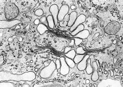
Additional info: Electron microscopy is essential for visualizing organelles, viruses, and macromolecular complexes.
Cell Types and Cellular Organization
Prokaryotic vs. Eukaryotic Cells
Cells are classified into two main types based on their internal organization: prokaryotic and eukaryotic.
Prokaryotic cells: Lack membrane-bound organelles and a nucleus. DNA is located in a region called the nucleoid. Examples include Bacteria and Archaea.
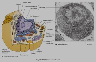
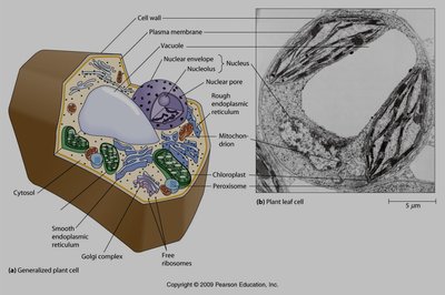
Eukaryotic cells: Have a true nucleus enclosed by a nuclear envelope and possess various membrane-bound organelles (e.g., mitochondria, endoplasmic reticulum, Golgi apparatus).
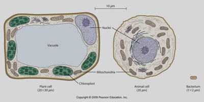
Typical Cells: Bacteria, Animal, and Plant Cells
Bacteria are prokaryotic cells, while animal and plant cells are eukaryotic. Each has unique features:
Bacteria: No membrane-bound organelles, no nuclear envelope, cell wall present.
Animal cells: No cell wall, contain lysosomes, small vacuoles, centrioles present, shape is round or irregular.
Plant cells: Have a cell wall, chloroplasts, large central vacuole, fixed shape, centrioles only in lower plants.
Feature | Plant Cell | Animal Cell |
|---|---|---|
Shape | Fixed | Round or irregular, can change |
Cilia, flagella | Very rare | Present |
Chloroplasts and other plastids | Yes | No |
Cell wall | Yes | No |
Lysosomes | Not found | Present in cytoplasm |
Vacuoles | One large | Small in cytoplasm |
Centrioles | Only in lower plants | Yes |
Units and Scale in Cell Biology
Metric Prefixes and Units
Understanding the scale of biological structures requires familiarity with metric prefixes and units commonly used in cell biology.
Micrometer (μm): 1 μm = meters
Nanometer (nm): 1 nm = meters
Common prefixes: kilo (k, ), milli (m, ), micro (μ, ), nano (n, )
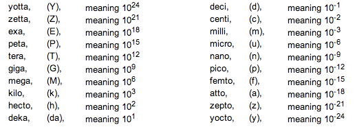
Additional info: Cells typically range from 1–100 μm in diameter; organelles and macromolecules are smaller.
Scale of Biological Structures
Biological structures span a wide range of sizes, from atoms to entire cells. Microscopes are essential for visualizing structures below the limit of unaided vision.
Light microscopes: Can resolve structures down to about 0.2 μm, such as cells and some organelles.
Electron microscopes: Can resolve structures as small as 0.2 nm, such as viruses, ribosomes, and macromolecules.
Summary Table: Plant Cell vs. Animal Cell
Feature | Plant Cell | Animal Cell |
|---|---|---|
Shape | Fixed | Round or irregular, can change |
Cilia, flagella | Very rare | Present |
Chloroplasts and other plastids | Yes | No |
Cell wall | Yes | No |
Lysosomes | Not found | Present in cytoplasm |
Vacuoles | One large | Small in cytoplasm |
Centrioles | Only in lower plants | Yes |
