 Back
Back15:Membrane Transport and Energy Conversion in Cell Biology
Study Guide - Smart Notes
 Tailored notes based on your materials, expanded with key definitions, examples, and context.
Membrane Transport: Transmitter-Gated Ion Channels and Synaptic Transmission
Transmitter-Gated Ion Channels at Chemical Synapses
Transmitter-gated ion channels are essential for converting chemical signals into electrical signals at chemical synapses, facilitating communication between neurons and their target cells.
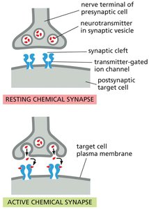
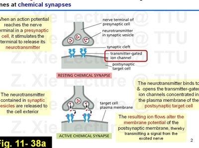
Chemical Synapse Structure: A chemical synapse consists of a presynaptic nerve terminal, synaptic vesicles containing neurotransmitter, a synaptic cleft, and a postsynaptic target cell membrane containing transmitter-gated ion channels.
Signal Transmission: When an action potential reaches the presynaptic terminal, it triggers the release of neurotransmitter into the synaptic cleft. The neurotransmitter binds to and opens transmitter-gated ion channels on the postsynaptic membrane, altering the membrane potential and transmitting the signal.
Resting vs. Active Synapse: In the resting state, ion channels are closed. Upon neurotransmitter binding, the channels open, allowing ion flow and signal propagation.
Example: At the neuromuscular junction, acetylcholine is released from the nerve terminal and binds to acetylcholine receptors on the muscle cell, initiating muscle contraction.
Ultrastructure of Synapses
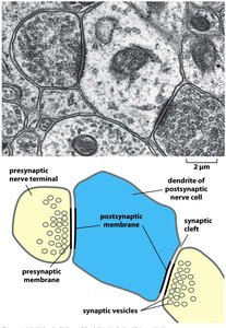
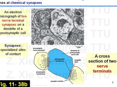
Electron micrographs reveal the specialized architecture of synapses, highlighting the close apposition of presynaptic and postsynaptic membranes and the presence of synaptic vesicles.
Specialized Contact: Synapses are specialized sites where nerve terminals closely approach the dendrites of postsynaptic cells, facilitating efficient neurotransmitter release and reception.
Visualization: Electron microscopy and schematic diagrams help illustrate the organization of synaptic components.
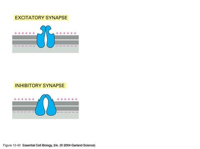
Excitatory and Inhibitory Synapses
Synapses can be classified as excitatory or inhibitory based on the effect of neurotransmitter release on the postsynaptic cell's membrane potential.
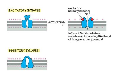
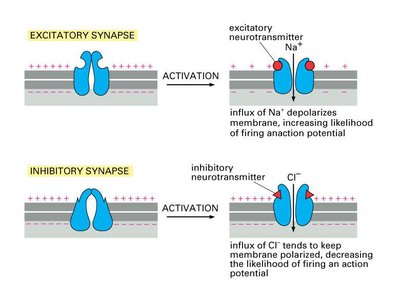
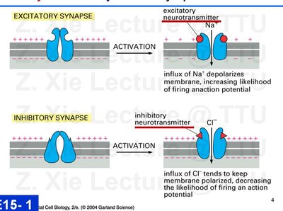
Excitatory Synapses: Activation leads to depolarization of the postsynaptic membrane, increasing the likelihood of firing an action potential. This is typically mediated by the influx of Na+ ions.
Inhibitory Synapses: Activation leads to hyperpolarization or stabilization of the membrane potential, decreasing the likelihood of firing an action potential. This is typically mediated by the influx of Cl- ions.
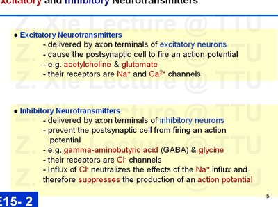
Excitatory and Inhibitory Neurotransmitters
Neurotransmitters are classified based on their effect on the postsynaptic cell. Their receptors are ion channels selective for specific ions.
Excitatory Neurotransmitters: Delivered by axon terminals of excitatory neurons, cause the postsynaptic cell to fire an action potential. Examples include acetylcholine and glutamate. Their receptors are Na+ and Ca2+ channels.
Inhibitory Neurotransmitters: Delivered by axon terminals of inhibitory neurons, prevent the postsynaptic cell from firing an action potential. Examples include gamma-aminobutyric acid (GABA) and glycine. Their receptors are Cl- channels. Influx of Cl- neutralizes the effects of Na+ influx, suppressing action potential generation.
Neuromuscular Junction and Acetylcholine Receptors
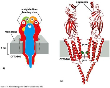
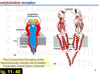
Structure and Function of Acetylcholine Receptors
The acetylcholine receptor at the neuromuscular junction is a classic example of an excitatory transmitter-gated cation channel, essential for muscle contraction.
Receptor Structure: The receptor is composed of multiple subunits forming a central pore that opens upon acetylcholine binding, allowing Na+ and K+ ions to flow.
Function: Activation of these receptors depolarizes the muscle cell membrane, triggering an action potential and subsequent muscle contraction.
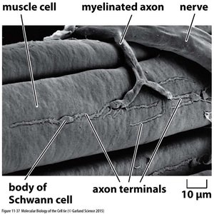
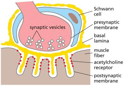
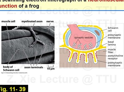
Neuromuscular Junction Ultrastructure
The neuromuscular junction is a specialized synapse between a motor neuron and a skeletal muscle fiber, facilitating rapid and efficient signal transmission for muscle contraction.
Key Components: Includes the presynaptic nerve terminal, synaptic vesicles, Schwann cells, basal lamina, and postsynaptic muscle membrane with acetylcholine receptors.
Visualization: Scanning electron micrographs and schematic diagrams illustrate the arrangement of these components.
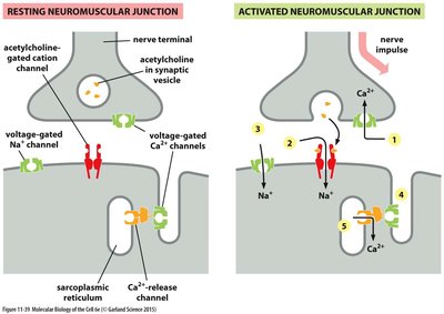
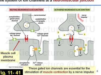
Ion Channels at the Neuromuscular Junction
Multiple types of ion channels coordinate to ensure efficient muscle contraction in response to nerve impulses.
Resting State: Ion channels are closed, and the muscle cell is at resting potential.
Activated State: Nerve impulse triggers the opening of voltage-gated Ca2+ channels, leading to acetylcholine release, which then opens acetylcholine-gated cation channels on the muscle membrane, followed by Na+ influx and muscle action potential.
Role of Sarcoplasmic Reticulum: Ca2+ release channels in the sarcoplasmic reticulum further amplify the contraction signal.
Pharmacological Modulation of Synaptic Transmission
Psychoactive Drugs and Synaptic Ion Channels
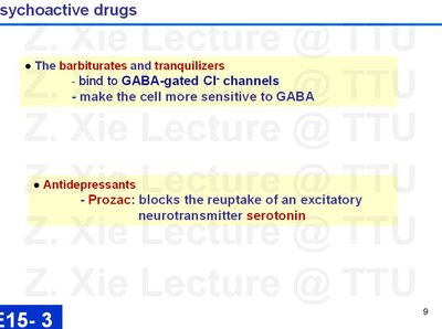
Many psychoactive drugs exert their effects by targeting transmitter-gated ion channels at synapses.
Barbiturates and Tranquilizers: Bind to GABA-gated Cl- channels, enhancing the inhibitory effect of GABA and making the cell more sensitive to inhibition.
Antidepressants (e.g., Prozac): Block the reuptake of the excitatory neurotransmitter serotonin, increasing its availability in the synaptic cleft and enhancing excitatory signaling.
Energy Conversion in Mitochondria and Chloroplasts
Overview of Chemiosmotic Energy Conversion
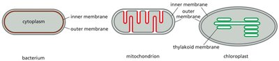
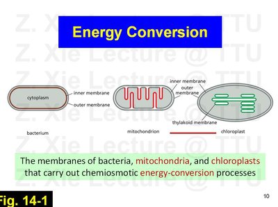
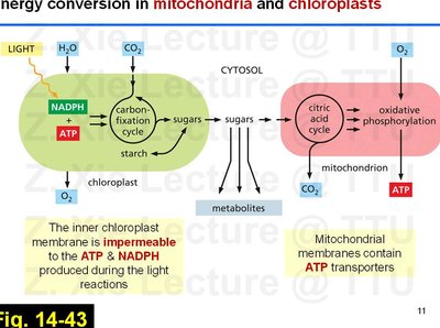
Energy conversion in cells is primarily carried out by mitochondria and chloroplasts, which use chemiosmotic mechanisms to generate ATP.
Membrane Systems: Bacteria, mitochondria, and chloroplasts possess specialized membranes that facilitate energy conversion processes.
ATP and NADPH Production: In chloroplasts, the inner membrane is impermeable to ATP and NADPH produced during the light reactions, while mitochondria contain ATP transporters to export ATP to the cytosol.
Electron Transport Chain and Proton Gradient
The electron transport chain (ETC) in mitochondria and chloroplasts establishes a proton gradient across the membrane, which is harnessed to synthesize ATP.
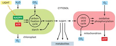
Mitochondria: Convert energy from chemical fuels (e.g., glucose) via the ETC, pumping protons across the inner membrane to generate a proton-motive force.
Chloroplasts: Use light energy to extract electrons from water, releasing O2 and generating a proton gradient across the thylakoid membrane.
Key Equation:
where is the change in free energy, is the number of electrons transferred, is the Faraday constant, and is the change in redox potential.
Stages of Chemiosmotic Coupling
Chemiosmotic coupling involves two main stages: generation of a proton gradient and synthesis of ATP by ATP synthase.
Stage 1: Energy from sunlight or food oxidation is used to pump protons across a membrane, creating an electrochemical gradient.
Stage 2: ATP synthase uses the energy stored in the proton gradient to drive the synthesis of ATP from ADP and inorganic phosphate.
ATP Synthesis Equation:
Summary Table: Comparison of Excitatory and Inhibitory Synapses
Type of Synapse | Neurotransmitter | Ion Channel | Effect on Membrane Potential | Result |
|---|---|---|---|---|
Excitatory | Acetylcholine, Glutamate | Na+, Ca2+ | Depolarization | Increased likelihood of action potential |
Inhibitory | GABA, Glycine | Cl- | Hyperpolarization or stabilization | Decreased likelihood of action potential |
Additional info: The notes above integrate and expand upon the provided lecture content, including definitions, mechanisms, and examples relevant to cell biology students. Images were included only when directly relevant to the adjacent explanation.
