 Back
BackMembranes: Structure, Function, and Chemistry
Study Guide - Smart Notes
 Tailored notes based on your materials, expanded with key definitions, examples, and context.
Chapter 7: Membranes – Their Structure, Function, and Chemistry
Learning Objectives
Describe the molecular structure of cell membranes
Explain the multiple functional roles that membranes play in the life of the cell
The Functions of Membranes
Overview of Membrane Functions
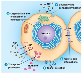
Biological membranes are essential for defining the boundaries of cells and their internal compartments. All biological membranes share a common structure: a fluid phospholipid bilayer embedded with a mosaic of proteins. This structure allows membranes to serve as selective barriers, regulate transport, and facilitate communication and adhesion between cells.
Permeability Barrier: The lipid component forms a hydrophobic barrier that restricts the free passage of water-soluble substances.
Transport Regulation: Membrane proteins control the movement of ions and molecules into and out of cells and organelles.
Signal Transduction: Membrane proteins detect and transmit external signals to the cell interior.
Cell Adhesion and Communication: Membranes mediate contact and adhesion between neighboring cells and participate in cell-to-cell communication.
Structural Support: Membranes interact with external structures such as the cell wall or extracellular matrix.
Models of Membrane Structure: An Experimental Perspective
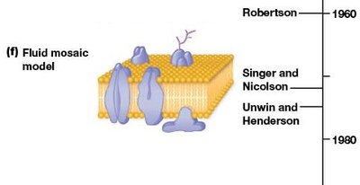
Historical Development of Membrane Models
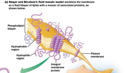
Our understanding of membrane structure has evolved over more than a century. Early models recognized the importance of lipids, but it was the fluid mosaic model proposed by Singer and Nicolson that became the universally accepted description. This model describes the membrane as a fluid lipid bilayer with proteins floating within or on it, allowing for dynamic movement and functional diversity.
Fluid Mosaic Model: Proteins with varying affinities for the hydrophobic membrane interior float in and on a fluid lipid bilayer.
Lipid Rafts: Specialized microdomains enriched in certain lipids and proteins, involved in cell signaling and interactions.
Membrane Lipids: The "Fluid" Part of the Model
Types and Distribution of Membrane Lipids
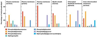
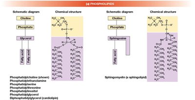
Membrane fluidity is primarily determined by its lipid composition. The main classes of membrane lipids are phospholipids, glycolipids, and sterols. The proportion of each lipid type varies depending on the membrane and organism.
Phospholipids: The most abundant membrane lipids, forming the basic bilayer structure.
Glycolipids: Lipids with carbohydrate groups, important for cell recognition.
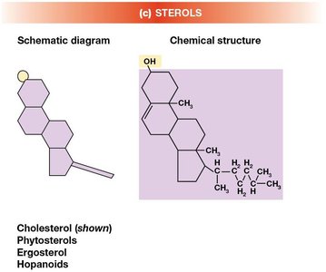
Sterols: Cholesterol in animal cells, phytosterols in plants, and hopanoids in some bacteria, which modulate membrane fluidity and stability.
Membrane Fluidity and Fatty Acid Composition
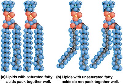
Membrane fluidity is critical for proper function and is influenced by the length and degree of unsaturation of fatty acid chains, as well as the presence of sterols. Cells can adjust these properties to maintain optimal fluidity under different conditions.
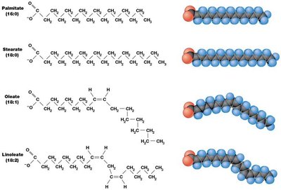
Long-chain fatty acids: Pack tightly, decreasing fluidity.
Unsaturated fatty acids: Contain cis double bonds that prevent tight packing, increasing fluidity.
Cholesterol: Modulates fluidity by preventing extremes of rigidity or fluidity.
Lipid Movement and Membrane Asymmetry
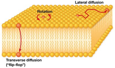
Phospholipids can move laterally within the membrane, rotate, and, rarely, flip-flop between layers. This movement contributes to membrane fluidity. Most membranes are asymmetric, with different lipid compositions on the inner and outer leaflets, contributing to functional specialization.
Lateral Diffusion: Rapid movement of lipids within the same layer.
Transverse Diffusion (Flip-Flop): Rare, requires enzymes called flippases.
Asymmetry: Different lipid types are distributed unequally between the two monolayers.
Membrane Proteins: The "Mosaic" Part of the Model
Classification of Membrane Proteins
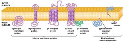
Proteins are major components of all cellular membranes and are classified based on their association with the lipid bilayer:
Integral Membrane Proteins: Span the membrane with hydrophobic regions embedded in the bilayer. Most have one or more α-helical transmembrane segments.
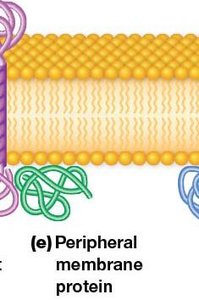
Peripheral Membrane Proteins: Hydrophilic proteins attached to the membrane surface by ionic and hydrogen bonds, often to phospholipid head groups.
Lipid-Anchored Proteins: Hydrophilic proteins covalently linked to lipid molecules embedded in the bilayer.
Functions of Membrane Proteins
Membrane proteins perform a wide variety of functions essential for cell survival and communication:
Enzymes: Catalyze specific reactions at the membrane surface.
Electron Carriers: Participate in electron transport chains.
Transport Proteins: Facilitate the movement of substances across the membrane.
Receptors: Bind signaling molecules such as hormones and neurotransmitters.
Cell Adhesion Molecules: Mediate cell-cell interactions and communication.
Structural Support: Stabilize and shape the membrane.
Many membrane proteins are glycoproteins, with carbohydrate side chains that serve as recognition markers on the cell surface.
Advances in Membrane Protein Study
Modern techniques such as X-ray crystallography, affinity labeling, and the use of specific antibodies have greatly advanced our understanding of membrane protein structure and function, which were previously difficult to study due to their amphipathic nature.
Summary Table: Types of Membrane Proteins
Type | Association with Membrane | Key Features | Examples |
|---|---|---|---|
Integral | Embedded within bilayer | Hydrophobic transmembrane segments | Ion channels, receptors |
Peripheral | Surface attachment | Hydrophilic, attached by non-covalent bonds | Signal transduction proteins |
Lipid-Anchored | Covalently linked to lipids | Hydrophilic protein, lipid anchor embedded | GPI-anchored proteins |
Key Equations and Concepts
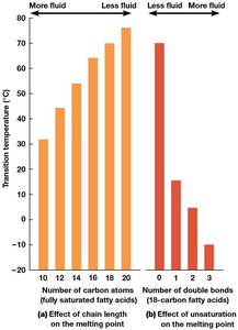
Membrane Fluidity: Increases with shorter fatty acid chains and more double bonds (unsaturation).
Transition Temperature (Tm): The temperature at which the membrane transitions from a rigid to a fluid state. Influenced by fatty acid composition and cholesterol content.
Additional info: The transition temperature is higher for membranes with longer, saturated fatty acids and lower for those with more unsaturated fatty acids or cholesterol.
