 Back
BackCh 7 - Membranes: Structure, Function, and Chemistry
Study Guide - Smart Notes
 Tailored notes based on your materials, expanded with key definitions, examples, and context.
Membrane Structure and Function
Overview of Biological Membranes
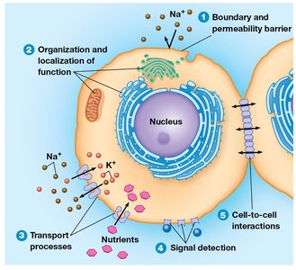
Biological membranes are essential structures that define the boundaries of cells and their internal compartments. All biological membranes share a common architecture: a fluid phospholipid bilayer embedded with a mosaic of proteins. This structure underlies the diverse functions that membranes perform in cellular life.
Boundary and Compartmentalization: Membranes separate the cell from its environment and partition the cell into functional compartments.
Selective Permeability: The lipid bilayer acts as a barrier to most water-soluble molecules, while membrane proteins regulate the transport of specific substances.
Signal Detection and Transduction: Membrane proteins detect external signals and initiate cellular responses.
Cell Adhesion and Communication: Membranes mediate contact and communication between neighboring cells and with the extracellular matrix.
Models of Membrane Structure: Historical Perspective
Development of Membrane Models
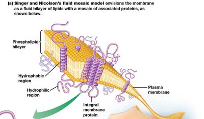
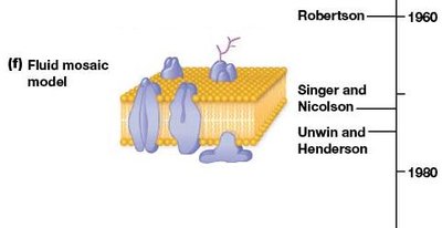
Our understanding of membrane structure has evolved through experimental studies over the past century. Early models recognized the importance of lipids, but the modern view is based on the fluid mosaic model proposed by Singer and Nicolson in 1972.
Lipid Bilayer Model: Gorter and Grendel (1925) demonstrated that membranes are composed of a lipid bilayer.
Davson-Danielli Model: Proposed that proteins coat the lipid bilayer on both sides.
Fluid Mosaic Model: Singer and Nicolson described membranes as a fluid lipid bilayer with proteins floating within or attached to it, allowing for lateral movement of components.
Membrane Lipids: The "Fluid" Part of the Model
Types and Distribution of Membrane Lipids
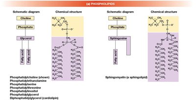
Membrane lipids are primarily phospholipids, glycolipids, and sterols. The proportion and types of lipids vary among different membranes and organisms.
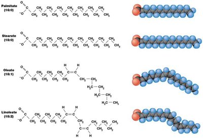
Phospholipids: The most abundant membrane lipids, consisting of a hydrophilic head and two hydrophobic fatty acid tails.
Glycolipids: Lipids with carbohydrate groups, important for cell recognition.
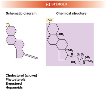
Sterols: Cholesterol in animal cells, phytosterols in plants, and hopanoids in some bacteria stabilize membrane structure.
Membrane Fluidity and Its Regulation
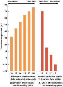
Membrane fluidity is crucial for proper function, affecting protein mobility and membrane permeability. Fluidity is influenced by the length and saturation of fatty acid chains and the presence of sterols.
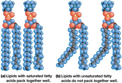
Fatty Acid Chain Length: Longer chains pack more tightly, decreasing fluidity.
Degree of Unsaturation: Unsaturated fatty acids (with cis double bonds) prevent tight packing, increasing fluidity.
Sterols: Cholesterol modulates fluidity by preventing tight packing at low temperatures and restricting movement at high temperatures.
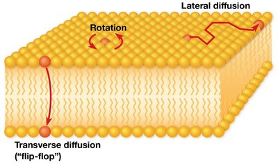
Lipid Movement and Membrane Asymmetry
Phospholipids can move laterally and rotate within the membrane, but "flip-flop" between layers is rare and requires enzymes called flippases. Membranes are asymmetric, with different lipid compositions on each side, contributing to functional specialization.
Lateral Diffusion: Rapid movement within the same leaflet.
Transverse Diffusion (Flip-Flop): Rare, catalyzed by flippases.
Asymmetry: Each leaflet has a distinct lipid composition, important for cell signaling and recognition.
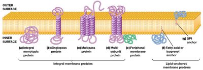
Membrane Proteins: The "Mosaic" Part of the Model
Classification of Membrane Proteins
Membrane proteins are classified based on their association with the lipid bilayer:
Integral Membrane Proteins: Span the bilayer with hydrophobic regions embedded in the membrane. Most are transmembrane proteins with one or more α-helical segments.
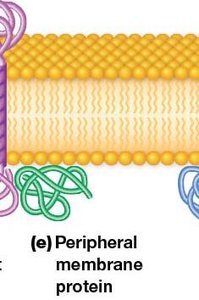
Peripheral Membrane Proteins: Hydrophilic proteins attached to the membrane surface by ionic or hydrogen bonds, often interacting with integral proteins or lipid head groups.
Lipid-Anchored Proteins: Covalently attached to lipids within the bilayer, but do not enter the hydrophobic core.
Functions of Membrane Proteins
Membrane proteins perform a wide range of functions essential for cell survival and communication:
Enzymatic Activity: Catalyze reactions at the membrane surface.
Transport: Facilitate movement of ions and molecules across the membrane.
Signal Transduction: Act as receptors for hormones and neurotransmitters.
Cell Recognition: Glycoproteins serve as markers for cellular identification.
Intercellular Joining: Mediate cell-cell adhesion and communication.
Attachment: Anchor the membrane to the cytoskeleton and extracellular matrix.
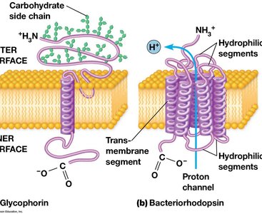
Glycoproteins and the Glycocalyx
Many membrane proteins are glycoproteins, with carbohydrate side chains exposed on the cell surface. These carbohydrates form the glycocalyx, which is involved in cell recognition, protection, and adhesion.
Summary Table: Types of Membrane Proteins
Type | Location | Association with Membrane | Example Function |
|---|---|---|---|
Integral | Spans bilayer | Hydrophobic regions embedded in core | Transporters, receptors |
Peripheral | Surface of membrane | Non-covalent interaction with lipids/proteins | Signal transduction, cytoskeletal attachment |
Lipid-anchored | Surface, covalently attached to lipid | Lipid anchor in bilayer | Enzymes, signaling proteins |
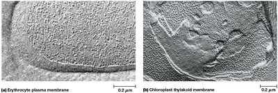
Experimental Advances in Membrane Biology
Modern Techniques
Recent advances in biochemistry, molecular biology, X-ray crystallography, affinity labeling, and antibody technology have greatly expanded our understanding of membrane protein structure and function, allowing for detailed analysis of their roles in cellular processes.
