Back
BackMembranes: Structure, Function, and Chemistry – Study Guide
Study Guide - Smart Notes
 Tailored notes based on your materials, expanded with key definitions, examples, and context.
Membranes: Structure, Function, and Chemistry
Functions of Biological Membranes
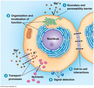
Biological membranes are essential for cellular life, providing structural boundaries and enabling specialized functions. They are composed of a lipid bilayer with embedded proteins, forming a dynamic and versatile interface between the cell and its environment.
Boundary and Permeability Barrier: Membranes separate the cell from its surroundings and regulate the passage of substances.
Organization and Localization of Function: Membranes compartmentalize cellular processes, allowing for efficient metabolic regulation.
Transport Processes: Membranes facilitate the movement of ions and nutrients via channels, carriers, and pumps.
Signal Detection: Membranes contain receptors that detect and respond to external signals.
Cell-to-Cell Interactions: Membranes mediate communication and adhesion between cells.
Models of Membrane Structure: Historical Perspective
The understanding of membrane structure has evolved through experimental observations and theoretical models.
Overton (1890s): Proposed a lipid coat based on the permeability of lipid-soluble substances.
Langmuir: Demonstrated that phospholipids are amphipathic, forming monolayers on water surfaces.
Gorter and Grendel (1925): Showed that cell membranes are lipid bilayers, with the surface area of extracted lipids being twice that of the cell surface.
Davson-Danielli: Suggested protein layers on either side of the lipid bilayer, but this model was inconsistent with protein properties.
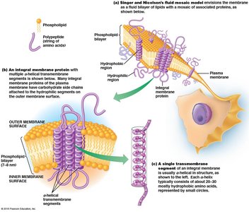
Singer and Nicholson (1972): Introduced the fluid mosaic model, describing membranes as a mosaic of proteins embedded in a fluid lipid bilayer.
Membrane Lipids: The "Fluid" Component
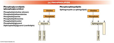
Phospholipids
Phospholipids are the most abundant lipids in membranes, forming the structural basis of the bilayer. They are amphipathic, with hydrophilic heads and hydrophobic tails.
Phosphoglycerides: Glycerol-based phospholipids with diverse R groups (e.g., phosphatidylcholine, phosphatidylethanolamine).
Phosphosphingolipids: Sphingosine-based phospholipids, such as sphingomyelin.
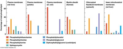
Phospholipid composition varies among membranes from different sources, reflecting functional specialization.
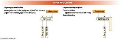
Glycolipids
Glycolipids are lipids with covalently attached carbohydrate groups, contributing to membrane asymmetry and cell recognition.
Glycoglycerolipids: Glycerol-based glycolipids.
Glycosphingolipids: Sphingosine-based glycolipids, including cerebrosides and gangliosides.
Sterols
Sterols are rigid, ring-structured lipids that modulate membrane fluidity and stability. Cholesterol is the principal sterol in animal cell membranes.
Cholesterol: Stabilizes membranes and prevents excessive fluidity.
Other sterols: Include campesterol, sitosterol, stigmasterol, ergosterol, and hopanoid (in prokaryotes).

Membrane Asymmetry
Membrane asymmetry refers to the unequal distribution of lipids between the two monolayers of the bilayer. Glycolipids are typically found in the outer layer, and asymmetry is established during membrane synthesis.
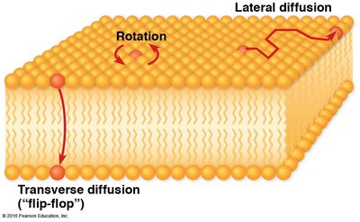
Lipid Mobility and Fluidity
Lipids move freely within their monolayer via rotation and lateral diffusion, contributing to membrane fluidity. Transverse diffusion (flip-flop) is rare and often catalyzed by specific proteins (flippases).
Rotation: Lipid molecules rotate around their axis.
Lateral diffusion: Lipids move sideways within the same layer.
Transverse diffusion: Lipids occasionally flip from one layer to the other.
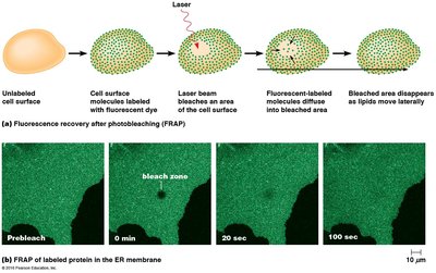
Measuring Lipid Mobility: FRAP
Fluorescence Recovery After Photobleaching (FRAP) is used to measure lipid mobility. Lipids labeled with fluorescent dye are bleached in a region, and recovery of fluorescence indicates lateral diffusion.
Factors Regulating Membrane Fluidity
Membrane fluidity is crucial for function and is regulated by several factors:
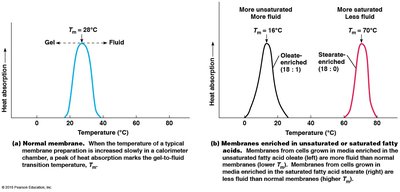
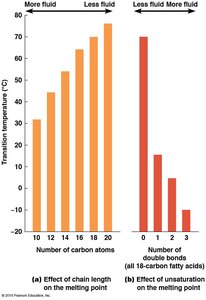
Temperature: Fluidity increases with temperature.
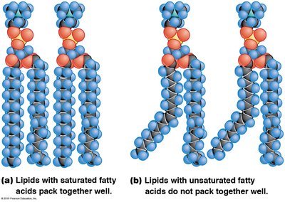
Fatty Acid Structure: Unsaturated fatty acids (with double bonds) increase fluidity; shorter hydrocarbon tails also increase fluidity.
Sterols: Cholesterol reduces fluidity at high temperatures and prevents gel formation at low temperatures.
Membrane Proteins: The "Mosaic" Component
Classes of Membrane Proteins
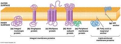
Membrane proteins are the main functional components of the membrane, classified by their association with the lipid bilayer:
Integral membrane proteins: Embedded within the bilayer, often spanning it with hydrophobic segments.
Peripheral proteins: Hydrophilic, attached to the membrane surface via non-covalent interactions.
Lipid-anchored proteins: Hydrophilic proteins covalently attached to lipid molecules within the bilayer.
Membrane-Spanning Structures
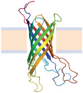
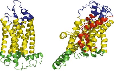
Integral membrane proteins often span the bilayer as α-helices or β-barrels, providing structural and functional diversity.
α-helix: Common in single-pass and multipass transmembrane proteins.
β-barrel: Found in some membrane channels and porins.
Protein Orientation and Asymmetry
Membrane proteins are asymmetrically oriented across the bilayer, with all molecules of a particular protein oriented the same way. Once inserted, proteins do not flip across the membrane.
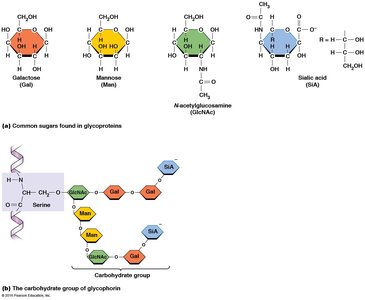
Glycosylation of Membrane Proteins
Many membrane proteins are glycosylated, meaning they have carbohydrate chains covalently attached. Glycosylation occurs in the endoplasmic reticulum (ER) and Golgi apparatus and is important for cell recognition and signaling.
N-linked glycosylation: Carbohydrate attached to the nitrogen atom of asparagine.
O-linked glycosylation: Carbohydrate attached to the oxygen atom of serine, threonine, or modified lysine/proline.
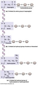
Carbohydrate chains can be straight or branched, ranging from 2 to 60 sugar units. Common sugars include galactose, mannose, N-acetylglucosamine, and sialic acid.