 Back
BackMembranes: Structure, Function, and Chemistry – Study Notes
Study Guide - Smart Notes
 Tailored notes based on your materials, expanded with key definitions, examples, and context.
Membranes: Their Structure, Function, and Chemistry
The Functions of Membranes
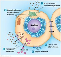
Biological membranes are essential for maintaining the integrity and functionality of cells. They serve as selective barriers, organize cellular processes, and facilitate communication and transport.
Boundary and Permeability Barrier: Membranes separate the cell from its environment and regulate the entry and exit of substances.
Organization and Localization of Function: Membranes compartmentalize cellular activities, allowing specialized functions in different organelles.
Transport Processes: Membranes contain proteins that mediate the movement of ions and molecules across the bilayer.
Signal Detection: Membranes possess receptors that detect and respond to external signals.
Cell-to-Cell Interactions: Membranes facilitate communication and adhesion between cells.
Models of Membrane Structure: An Experimental Approach
Historical Models of Membrane Structure
The understanding of membrane structure has evolved through experimental observations and theoretical models.
Overton's Lipid Hypothesis: Overton observed that lipid-soluble substances penetrate cells easily, suggesting a lipid-rich cell surface.
Langmuir's Monolayer Experiments: Langmuir demonstrated that phospholipids form a single molecular layer on water, indicating their amphipathic nature.
Gorter and Grendel's Bilayer Model: By extracting lipids from red blood cells, they found that the surface area of the lipid film was twice that of the cells, supporting the existence of a lipid bilayer.
Davson-Danielli Model: Proposed that membranes are lipid bilayers coated with protein layers on both sides, explaining certain permeability properties.
Problems with the Davson-Danielli Model: The discovery of globular, water-insoluble proteins and variable protein:lipid ratios challenged this model.
Singer and Nicholson's Fluid Mosaic Model: This model describes membranes as a fluid lipid bilayer with a mosaic of proteins embedded or associated with it, accounting for previous inconsistencies.

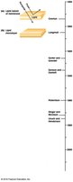
Experimental Timeline of Membrane Models
Year | Scientist(s) | Key Finding |
|---|---|---|
1890s | Overton | Lipid-rich cell surface |
Early 1900s | Langmuir | Phospholipid monolayer |
1925 | Gorter & Grendel | Lipid bilayer |
1935 | Davson & Danielli | Protein-lipid-protein sandwich |
1972 | Singer & Nicholson | Fluid mosaic model |
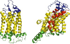
Unwin and Henderson: Transmembrane Segments
Most integral membrane proteins contain one or more hydrophobic segments that span the lipid bilayer, forming transmembrane domains essential for their function.
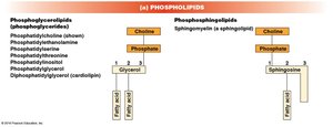
Membrane Lipids: The "Fluid" Part of the Model
Types of Membrane Lipids
Membranes are composed of various lipids that contribute to their fluidity and function.
Phospholipids: The most abundant membrane lipids, including phosphoglycerides and sphingolipids.
Glycolipids: Lipids with attached carbohydrate groups, such as cerebrosides and gangliosides.
Sterols: Cholesterol (in animals) and other sterols stabilize membrane structure.
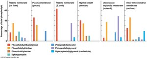


Membrane Asymmetry
Membrane asymmetry refers to the unequal distribution of lipids between the two monolayers of the bilayer. Glycolipids are typically found in the outer layer, and this asymmetry is established during membrane synthesis and maintained over time.
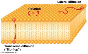
Lipid Mobility in Membranes
Lipids can move within their monolayer by rotation and lateral diffusion, which are rapid and random. Transverse diffusion (flip-flop) is rare due to the energetic barrier of moving the hydrophilic head through the hydrophobic core.
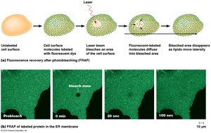
Measuring Lipid Mobility: FRAP
Fluorescence Recovery After Photobleaching (FRAP) is used to study lipid mobility. Lipids labeled with fluorescent dye are bleached in a small area, and the recovery of fluorescence indicates the rate of lipid movement.
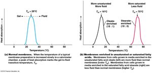
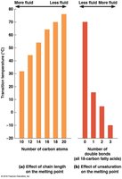
Factors Affecting Membrane Fluidity
Temperature: Higher temperatures increase membrane fluidity.
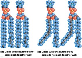
Fatty Acid Structure: Unsaturated fatty acids (with double bonds) increase fluidity; saturated fatty acids decrease it.
Hydrocarbon Tail Length: Shorter tails increase fluidity.
Sterol Content: Sterols reduce fluidity at high temperatures and prevent gelling at low temperatures.
Membrane Proteins: The "Mosaic" Part of the Model
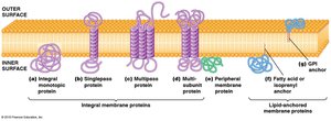
Classes of Membrane Proteins
Membrane proteins are classified based on their association with the lipid bilayer:
Integral Membrane Proteins: Embedded within the bilayer due to hydrophobic regions; may span the membrane (transmembrane proteins).
Peripheral Proteins: Hydrophilic proteins associated with the membrane surface.
Lipid-Anchored Proteins: Covalently attached to lipids within the bilayer but do not span the membrane.


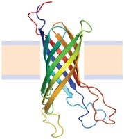
Membrane-Spanning Structures
Integral membrane proteins can span the bilayer as α-helices or β-barrels, providing structural diversity and functional specificity.
Asymmetric Orientation of Membrane Proteins
Membrane proteins are asymmetrically oriented in the bilayer, with all molecules of a particular protein oriented the same way. Once inserted, proteins do not flip-flop across the membrane.
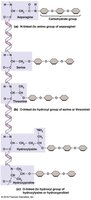
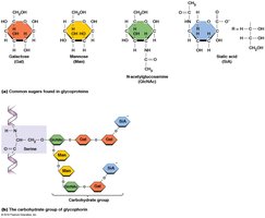
Glycosylation of Membrane Proteins
Many membrane proteins are glycosylated, meaning they have carbohydrate chains covalently attached. Glycosylation occurs in the endoplasmic reticulum (ER) and Golgi apparatus and can be N-linked (to asparagine) or O-linked (to serine, threonine, or modified residues).
Summary Table: Major Components of Biological Membranes
Component | Function | Example |
|---|---|---|
Phospholipids | Main structural framework | Phosphatidylcholine |
Glycolipids | Cell recognition, protection | Gangliosides |
Sterols | Membrane stability | Cholesterol |
Integral Proteins | Transport, signaling | Ion channels |
Peripheral Proteins | Support, signaling | Cytoskeletal anchors |
Lipid-Anchored Proteins | Enzyme activity, signaling | GPI-anchored proteins |
