 Back
Signal Transduction Mechanisms: Electrical and Synaptic Signaling in Neurons
Study Guide - Smart Notes
 Tailored notes based on your materials, expanded with key definitions, examples, and context.
Signal Transduction in Neurons
Electrical Signaling and Reflex Circuits
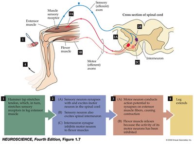
Neurons communicate via electrical signals, which are essential for rapid information transfer in the nervous system. A classic example is the knee-jerk reflex, which demonstrates the basic principles of neuronal signaling and synaptic transmission.
Reflex Circuit: The knee-jerk response involves sensory neurons, interneurons, and motor neurons, illustrating the flow of information from stimulus to response.
Steps: Sensory input activates a circuit that leads to muscle contraction and leg extension.
Application: Reflexes are used clinically to assess nervous system function.
Membrane Potential
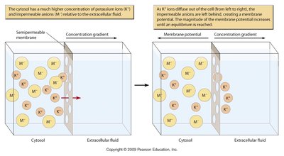
The membrane potential is the voltage difference across a cell's plasma membrane, determined by ion concentration gradients and membrane permeability. It is fundamental to neuronal excitability and signal transmission.
Key Factors: Ion concentration gradients (Na+, K+, Cl-) and selective permeability of the membrane.
Resting Membrane Potential: Typically negative inside the cell due to high K+ inside and high Na+ outside.
Equation: The Nernst equation calculates the equilibrium potential for each ion:

Types of Membrane Potential Changes
Neurons exhibit several types of membrane potential changes, each with distinct roles in signaling.
Depolarization: Membrane potential becomes less negative.
Repolarization: Return to resting potential after depolarization.
Hyperpolarization: Membrane potential becomes more negative than resting.
Functional Types: Receptor potential, synaptic potential, action potential.
Action Potential
An action potential is a rapid, transient change in membrane potential that propagates along the axon, enabling communication between neurons.
Phases: Depolarization (Na+ influx), repolarization (K+ efflux), and hyperpolarization.
Ion Channels: Voltage-gated Na+ and K+ channels are critical for action potential generation.
Sequence: Opening of Na+ channels causes rapid depolarization, followed by opening of K+ channels for repolarization.
Equation: Hodgkin-Huxley model describes the ionic basis of action potentials:

Synaptic Transmission
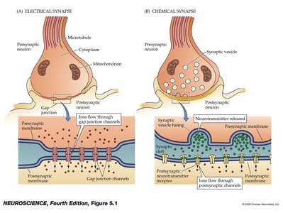
Synaptic transmission is the process by which neurons communicate with each other or with effector cells. It can be electrical or chemical.
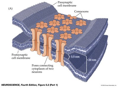
Electrical Synapses: Direct flow of ions through gap junctions, allowing rapid transmission.
Chemical Synapses: Neurotransmitter release from presynaptic neuron activates receptors on postsynaptic cell.
Sequence: Action potential arrives, Ca2+ influx triggers neurotransmitter release, postsynaptic response occurs.
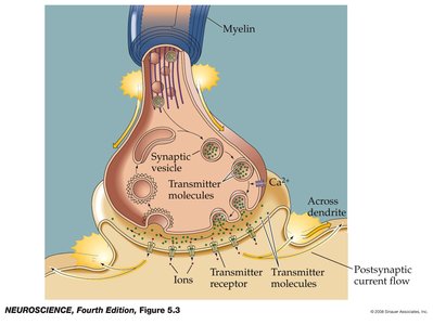
Criteria for Neurotransmitters
Neurotransmitters are defined by specific criteria that ensure their role in synaptic communication.
Presence: Must be present in the presynaptic neuron.
Release: Must be released in response to presynaptic depolarization, and release must be calcium-dependent.
Receptors: Specific receptors for the substance must be present on the postsynaptic cell.
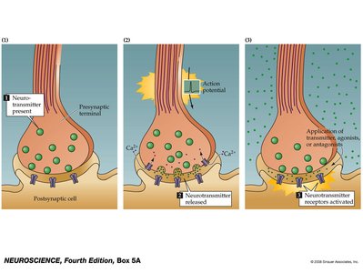
Molecular Signaling Mechanisms
Classes of Cell Signaling Molecules
Cell signaling molecules are classified based on their distance of action and chemical properties.
Synaptic: Neurotransmitters act locally at synapses.
Paracrine: Molecules act on nearby cells.
Endocrine: Hormones act on distant cells via the bloodstream.
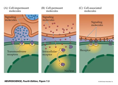
Receptors and Secondary Messenger Systems
Receptors are proteins that detect signaling molecules and initiate cellular responses. Secondary messengers amplify and propagate the signal within the cell.
Types of Receptors: Channel-linked, enzyme-linked, G protein-coupled, and intracellular receptors.
Secondary Messengers: Molecules such as cAMP, Ca2+, and IP3 rapidly transmit signals inside the cell.
Amplification: Secondary messengers allow a single extracellular signal to produce a large intracellular response.
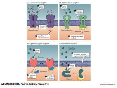
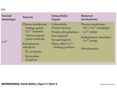
Calcium Signaling and Homeostasis
Calcium ions (Ca2+) serve as a universal secondary messenger in many signaling pathways. Their concentration is tightly regulated by various cellular mechanisms.
Sources: Voltage-gated Ca2+ channels, ligand-gated channels, endoplasmic reticulum.
Targets: Calmodulin, protein kinases, ion channels, synaptotagmin.
Removal: Ca2+ pumps, Na+/Ca2+ exchangers, mitochondria.


Summary Table: Key Signaling Pathways
The following table summarizes important signaling pathways, their sources, targets, and steps involved:
Pathway | Source | Targets | Steps |
|---|---|---|---|
NO Signaling | NO synthase, from arginine | Smooth muscle (relaxation) | Guanylyl cyclase (cGMP), PKG phosphorylation |
Protein Kinases | Membrane or cytoplasm, ATP | Platelets, cell growth, ion channels, signaling cascades | Autophosphorylation, target phosphorylation |
Hormonal Signaling | Hormone-secreting cells | Hormone receptors (membrane or intracellular) | Receptor activation, IP3, Ca2+, kinase activation |
Growth Factor Signaling | Developing or injured tissues | Gene expression | Receptor, kinases, signaling proteins translocation |
Apoptosis | Hormones or cell damage signals | DNAases, flippase, pro-apoptotic proteins | Death receptor, caspases, mitochondria, Ca2+ |
Additional info: Academic context was added to clarify the mechanisms and significance of each pathway.
