 Back
BackSignal Transduction Mechanisms: Electrical and Synaptic Signaling in Neurons
Study Guide - Smart Notes
 Tailored notes based on your materials, expanded with key definitions, examples, and context.
Signal Transduction Mechanisms: Electrical and Synaptic Signaling in Neurons
Overview of Neuronal Signaling
Neurons are specialized cells of the nervous system that transmit information via electrical and chemical signals. The regulation of ion flow across cell membranes is fundamental to neuronal function, enabling the generation and propagation of nerve impulses over long distances.
Cells of the Nervous System
Neurons: Cells that send and receive electrical impulses (nerve impulses).
Glial Cells: Supportive cells with various functions, including immune defense (microglia), myelin sheath formation (oligodendrocytes in the CNS, Schwann cells in the PNS), and regulation of the extracellular environment (astrocytes).
Types of Neurons
Sensory Neurons: Detect external or internal stimuli.
Motor Neurons: Transmit signals from the central nervous system (CNS) to muscles and glands.
Interneurons: Process and relay information within the CNS.
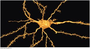
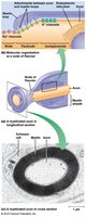
Neuron Structure and Function
Neurons have a unique structure adapted for signal transmission:
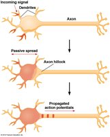
Cell Body (Soma): Contains the nucleus and organelles.
Dendrites: Receive incoming signals from other neurons.
Axon: Conducts electrical impulses away from the cell body.
Axon Hillock: Site where action potentials are initiated.
Synapse: Junction where information is transmitted to another cell.
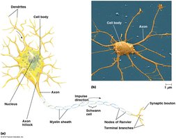
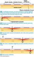
Myelin Sheath and Nodes of Ranvier
Myelin Sheath: Insulating layer formed by oligodendrocytes (CNS) or Schwann cells (PNS) that increases the speed of impulse conduction.
Nodes of Ranvier: Gaps in the myelin sheath where action potentials are regenerated.
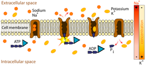
Membrane Potential and Ion Distribution
Establishing the Resting Potential
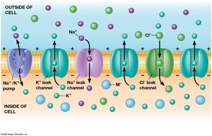
The resting membrane potential is the voltage difference across the plasma membrane of a neuron at rest, typically around –60 mV. This potential is established by the differential distribution of ions, primarily potassium (K+), sodium (Na+), and chloride (Cl–), across the membrane.
Sodium-Potassium Pump (Na+/K+ ATPase): Actively transports 3 Na+ ions out and 2 K+ ions into the cell, maintaining concentration gradients.
Ion Channels: Allow passive movement of ions down their concentration gradients (leak channels for K+, Na+, and Cl–).
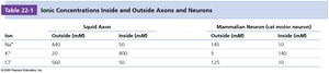
Ionic Concentrations Inside and Outside Neurons
Ion | Outside (mM) | Inside (mM) |
|---|---|---|
Na+ | 145 | 10 |
K+ | 5 | 140 |
Cl– | 110 | 4 |
Ion Channel Function
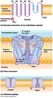
Ion channels are selective for specific ions and can be gated by voltage or ligands. The opening and closing of these channels underlie changes in membrane potential.
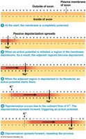
Action Potentials
Electrical Excitability and Action Potentials
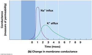
Neurons are electrically excitable cells that respond to stimuli with action potentials—rapid, transient changes in membrane potential. Action potentials are generated by voltage-gated Na+ and K+ channels.
Depolarization: Membrane potential becomes less negative due to Na+ influx.
Repolarization: Return to resting potential due to K+ efflux.
Hyperpolarization (Undershoot): Membrane potential temporarily becomes more negative than resting potential.
Phases of the Action Potential
Resting State: Most voltage-gated channels are closed.
Depolarizing Phase: Na+ channels open, Na+ enters the cell.
Repolarizing Phase: Na+ channels inactivate, K+ channels open, K+ leaves the cell.
Hyperpolarizing Phase: K+ channels remain open, membrane potential drops below resting value.

Propagation of Action Potentials
Action potentials are propagated along the axon without loss of strength. Depolarization at one region triggers depolarization in adjacent regions, ensuring unidirectional signal transmission.
Myelination and Saltatory Conduction
Myelinated axons conduct action potentials more rapidly via saltatory conduction, where the action potential jumps from one node of Ranvier to the next.
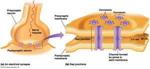
Synaptic Transmission
Types of Synapses
Electrical Synapses: Direct cytoplasmic connections via gap junctions allow rapid ion flow between neurons.
Chemical Synapses: Presynaptic and postsynaptic cells are separated by a synaptic cleft; neurotransmitters mediate signal transmission.
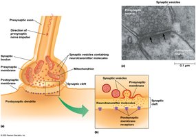
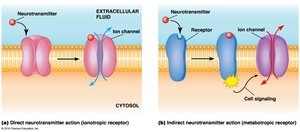
Neurotransmitter Receptors
Ionotropic Receptors: Ligand-gated ion channels that mediate rapid responses.
Metabotropic Receptors: G-protein-coupled receptors that initiate slower, indirect signaling cascades.
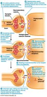
Neurotransmitter Release and Inactivation
Release: Action potentials trigger Ca2+ influx, leading to vesicle fusion and neurotransmitter release into the synaptic cleft.
Inactivation: Neurotransmitters are rapidly removed by enzymatic degradation (e.g., acetylcholinesterase) or reuptake into cells.
Examples of Neurotransmitters
Acetylcholine: Excitatory at cholinergic synapses.
Catecholamines: Dopamine, norepinephrine, epinephrine (adrenergic synapses).
Amino Acids: Glutamate (excitatory), GABA and glycine (inhibitory).
Neuropeptides, gases, and lipids also function as neurotransmitters.
Agonists and Antagonists
Agonists: Compounds that activate neurotransmitter receptors.
Antagonists: Compounds that block neurotransmitter receptors.
Summary Table: Key Features of Neuronal Signaling
Feature | Description |
|---|---|
Resting Potential | Voltage across membrane at rest (~–60 mV) |
Action Potential | Rapid depolarization and repolarization of membrane |
Myelin Sheath | Insulates axons, speeds conduction |
Synapse | Junction for signal transmission (electrical or chemical) |
Neurotransmitter | Chemical messenger released at synapse |
Additional info: This summary integrates foundational concepts from cell biology and neurobiology, providing a comprehensive overview suitable for exam preparation in a college-level cell biology course.
