 Back
BackSignal Transduction Mechanisms: Messengers and Receptors
Study Guide - Smart Notes
 Tailored notes based on your materials, expanded with key definitions, examples, and context.
Signal Transduction Mechanisms: Messengers and Receptors
Overview of Cell Signaling
Cell signaling is a fundamental process by which cells communicate with each other to coordinate their activities. This communication is achieved through the transmission of regulatory chemical messengers, which can be displayed on cell surfaces or released into the extracellular environment. - Intercellular Communication: Cells produce signals to influence the behavior of other cells, either locally or at a distance. - Multicellular Coordination: Specialized cells in multicellular organisms are regulated by chemical messengers.
Classification of Signaling Molecules
Signaling molecules are categorized based on the distance between their site of production and their target cells. - Endocrine signals: Produced far from target tissues and transported via the bloodstream. - Paracrine signals: Diffusible and act over short distances. - Juxtacrine signals: Require direct physical contact between cells. - Autocrine signals: Act on the same cell that produces them. 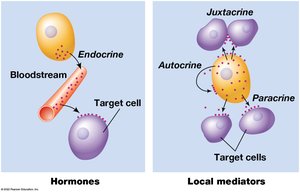
Receptors and Ligands
When a chemical messenger reaches its target cell, it binds to specific receptors, either on the cell surface or inside the cell. - Ligand: A molecule that binds to a receptor to initiate a cellular response. - Receptor: A protein that recognizes and binds the ligand, triggering downstream signaling events.
Information Flow During Cell Signaling
The binding of a ligand to its receptor initiates a cascade of intracellular events, often involving second messengers, which ultimately lead to changes in cellular behavior or gene expression. 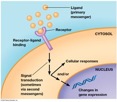
Signal Transduction
Signal transduction refers to the process by which cells convert an extracellular signal (ligand binding) into a functional response. - Second Messengers: Molecules or ions produced within the cell in response to receptor activation. - Cascade Effect: A series of biochemical changes that amplify the signal and produce a cellular response.
Quantitative Interactions: Receptor Binding
The interaction between ligands and receptors is highly specific and can be described quantitatively. - Affinity: The strength of the binding between a ligand and its receptor. - Saturation: Occurs when most receptors are occupied by ligands, typically at high ligand concentrations. - Binding Curve: The relationship between ligand concentration and receptor occupancy can be represented mathematically. where is ligand concentration and is the dissociation constant. 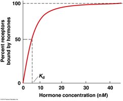
Agonists and Antagonists
Synthetic ligands can be designed to modulate receptor activity. - Agonists: Activate the receptor, mimicking the natural ligand. - Antagonists: Bind the receptor without activating it, blocking the natural ligand.
Signal Amplification
Cells can amplify signals received from very small quantities of ligand. - Amplification: Each step in the signaling cascade increases the number of activated molecules, resulting in a large cellular response from a small initial signal. 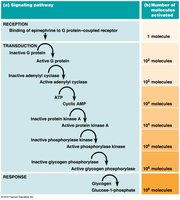
Types of Receptors and Signal Transduction Pathways
Cells utilize a limited number of basic signaling pathways, each involving specific types of ligands and receptors. - Hydrophilic ligands: Bind to transmembrane receptor proteins. - Hydrophobic ligands: Act on cytosolic or nuclear receptors. 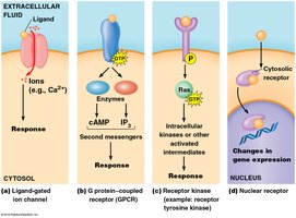
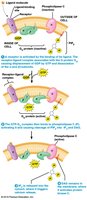
G Protein–Coupled Receptors (GPCRs)
Structure and Function of GPCRs
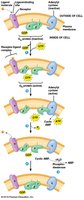
GPCRs are a major class of membrane receptors characterized by seven transmembrane helices. - Activation: Ligand binding induces a conformational change that activates a G protein. 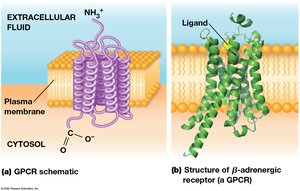
Activation and Inactivation of G Proteins
G proteins act as molecular switches, cycling between "on" (GTP-bound) and "off" (GDP-bound) states. - Heterotrimeric G proteins: Composed of Gα, Gβ, and Gγ subunits. - Activation: Ligand binding causes Gα to exchange GDP for GTP and dissociate from Gβγ. - Inactivation: Gα hydrolyzes GTP to GDP and reassociates with Gβγ. 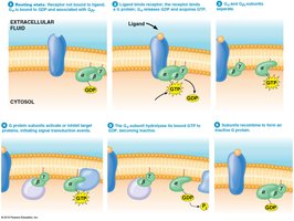
Signaling by βγ Subunits
In some pathways, the βγ subunit of the G protein initiates signal transduction, such as opening potassium channels in response to acetylcholine. 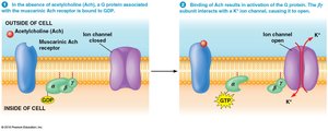
Second Messengers: Cyclic AMP and Calcium
Cyclic AMP (cAMP) as a Second Messenger
cAMP is a key second messenger produced from ATP by adenylyl cyclase, which is activated by Gα subunits. - Function: cAMP activates protein kinase A (PKA), which phosphorylates target proteins. 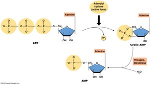
cAMP Function and Protein Kinase A Activation
- PKA Structure: Composed of regulatory and catalytic subunits. - Activation: cAMP binds regulatory subunits, releasing active catalytic subunits. 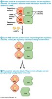
Inositol Trisphosphate (IP3) and Diacylglycerol (DAG)
IP3 and DAG are second messengers generated from PIP2 by phospholipase C. - IP3: Diffuses through the cytosol and opens calcium channels in the ER. - DAG: Activates protein kinase C (PKC). 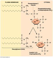
Calcium Signaling
Calcium Release and Regulation
Calcium ions (Ca2+) are essential for many cellular functions. - Calcium ATPases: Maintain low cytosolic calcium by pumping Ca2+ out of the cell or into the ER. - Sodium-calcium exchangers: Further reduce cytosolic calcium. - Mitochondria: Transport calcium into the matrix. 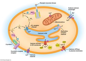
Calcium-Induced Calcium Release
- IP3 receptor channel: Releases calcium from ER. - Ryanodine receptor channel: Opens in response to increased cytosolic calcium, amplifying the signal.
Calmodulin and Effector Proteins
Calmodulin is a calcium-binding protein that mediates many calcium-activated processes. - Structure: Each calmodulin molecule binds four Ca2+ ions, changing conformation to activate target proteins. 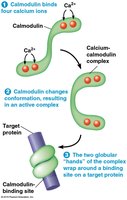
Protein Kinase-Associated Receptors
Receptor Tyrosine Kinases (RTKs)
RTKs are membrane receptors with intrinsic kinase activity, often activated by growth factors such as insulin, FGF, EGF, and NGF. - Structure: Single polypeptide chain with extracellular ligand-binding domain and cytosolic tyrosine kinase domain. 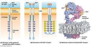
Activation and Signal Transduction Cascade
- Dimerization and Autophosphorylation: Ligand binding causes receptor dimerization and phosphorylation of tyrosine residues. - SH2 Domain: Cytosolic proteins with SH2 domains bind phosphotyrosine residues, initiating downstream signaling. 
Ras and MAP Kinase Pathway
Ras is a small G protein that regulates cell proliferation. - Activation: Ras is activated by GEF (Sos) via GRB2 binding to RTK. - MAP Kinase Cascade: Activated Ras triggers phosphorylation of Raf, MEK, and MAPKs, which regulate gene expression. 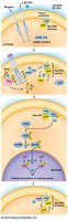


Inactivation of Ras
Ras is inactivated by GTPase activating proteins (GAPs), which facilitate GTP hydrolysis. 
Dominant Negative and Constitutively Active Mutations
- Dominant Negative Mutations: Mutant receptors dimerize with normal receptors, blocking function. 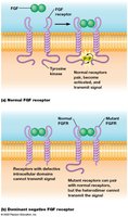 - Constitutively Active Mutations: Mutations keep receptors active without ligand, leading to diseases such as achondroplasia and cancer.
Signal Integration and Crosstalk
Integration of Signaling Pathways
Cells coordinate responses to multiple signals through scaffolding proteins and pathway integration. - Scaffolding Proteins: Recruit kinases into complexes for efficient signaling. 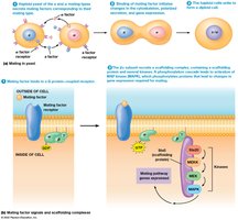
Signaling Crosstalk
Multiple pathways can converge or influence each other, forming a network of biochemical interactions. 
Hormones and Steroid Signaling
Hormone Categories
Endocrine hormones are classified as: - Amino acid derivatives: e.g., epinephrine - Peptides: e.g., vasopressin - Proteins: e.g., insulin - Lipid-like hormones: e.g., steroids
Steroid Hormones
Steroid hormones are hydrophobic and bind cytosolic receptors, which then translocate to the nucleus to regulate gene expression. 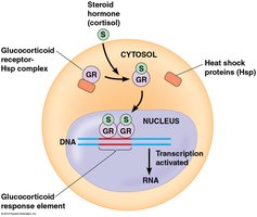
Gaseous Signals
Dissolved gases such as O2, CO2, and nitric oxide (NO) can act as cell signals, with NO playing a key role in nervous system signaling.
Summary Table: Types of Cell Signaling
Type | Distance | Example |
|---|---|---|
Endocrine | Long-range | Hormones (insulin) |
Paracrine | Short-range | Growth factors |
Juxtacrine | Direct contact | Immune cell signaling |
Autocrine | Self-signaling | Cancer cell growth |
Key Equations
Receptor-Ligand Binding: Signal Amplification: Each step in a cascade multiplies the number of activated molecules, leading to a large response from a small initial signal.
Conclusion
Signal transduction mechanisms are essential for cellular communication, integrating a variety of messengers, receptors, and pathways to regulate cell behavior and gene expression. Understanding these processes is fundamental to cell biology and has broad implications for physiology, development, and disease.
