 Back
BackSignal Transduction via Gq Proteins, IP3/DAG Pathway, and Calcium Regulation in Cells
Study Guide - Smart Notes
 Tailored notes based on your materials, expanded with key definitions, examples, and context.
Signal Transduction Mechanisms: Gq Proteins, IP3/DAG Pathway, and Calcium Regulation
Overview of G Proteins and Gq Subtype
G proteins are heterotrimeric proteins that play a central role in transmitting signals from cell surface receptors to intracellular effectors. The Gq subtype is particularly important in activating phospholipase C and initiating the IP3/DAG signaling pathway.
Structure: G proteins consist of three subunits: alpha (α), beta (β), and gamma (γ).
Activation: Upon receptor stimulation, GDP on the α subunit is replaced by GTP, causing the α subunit to dissociate and interact with target proteins.
Gq Function: The Gq α subunit activates phospholipase C (PLC), which is crucial for the generation of second messengers.
IP3 and DAG: Second Messengers in Signal Transduction
Inositol trisphosphate (IP3) and diacylglycerol (DAG) are key second messengers produced by the cleavage of phosphatidylinositol 4,5-bisphosphate (PIP2) in the plasma membrane. They mediate various cellular responses by mobilizing calcium and activating protein kinase C, respectively.
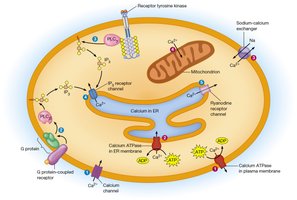
Production: Ligand binding to a G protein-coupled receptor (GPCR) activates Gq, which in turn activates PLC. PLC cleaves PIP2 into IP3 (water-soluble) and DAG (membrane-bound).
IP3: Diffuses through the cytosol and binds to IP3 receptors on the endoplasmic reticulum (ER), triggering the release of Ca2+ into the cytosol.
DAG: Remains in the membrane and activates protein kinase C (PKC), which regulates processes such as cell growth, ion channel activity, cytoskeletal changes, and protein secretion.

Calcium Release and Its Regulation
Calcium ions (Ca2+) serve as a universal second messenger in cells, regulating a wide range of cellular functions. The release of Ca2+ from intracellular stores is tightly controlled by IP3 and other signaling pathways.
IP3-Mediated Calcium Release: IP3 binds to ligand-gated calcium channels on the ER, causing them to open and release Ca2+ into the cytosol.
Regulation Mechanisms: Calcium levels are regulated by pumps (e.g., Ca2+-ATPases), exchangers, and buffering proteins to maintain cellular homeostasis.
Measurement and Manipulation: Calcium indicators (dyes or proteins) and ionophores (e.g., ionomycin) are used experimentally to study calcium dynamics.
Physiological Roles of Calcium
Calcium ions are essential for numerous cellular processes, including muscle contraction, neuronal signaling, cell migration, and growth. Dysregulation of calcium can lead to cellular dysfunction and death.
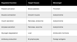
Examples of Calcium-Regulated Functions:
Regulated Function | Target Tissues | Messenger |
|---|---|---|
Platelet activation | Blood platelets | Thrombin |
Muscle contraction | Smooth muscle | Acetylcholine |
Insulin secretion | Pancreas, endocrine | Acetylcholine |
Amylase secretion | Pancreas, exocrine | Acetylcholine |
Glycogen degradation | Liver | Antidiuretic hormone |
Antibody production | B lymphocytes | Foreign antigens |
Calmodulin: A Key Calcium Sensor
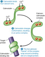
Calmodulin is a calcium-binding protein that mediates many of the effects of Ca2+ in the cell. Upon binding calcium, calmodulin undergoes a conformational change, enabling it to activate various target proteins, including kinases and phosphatases.
Structure: Calmodulin has two globular domains, each capable of binding two Ca2+ ions.
Activation: Binding of four Ca2+ ions induces a conformational change, allowing calmodulin to wrap around and activate target proteins.
Targets: Includes myosin light-chain kinase (for muscle contraction) and other enzymes involved in cellular regulation.
Consequences of Calcium Dysregulation
Proper regulation of intracellular calcium is critical for cell survival. If calcium is not regulated, proteins like calmodulin remain constantly active, leading to uncontrolled cellular activity and eventual cell death.
Cellular Effects: Persistent activation of calcium-dependent processes disrupts normal cell function and prevents the cell from resetting after stimulation.
Cell Death: Chronic dysregulation can trigger cell death pathways due to metabolic exhaustion and loss of homeostasis.
Summary Table: Key Steps in Gq/IP3/DAG/Calcium Pathway
Step | Description |
|---|---|
1. Ligand binds GPCR | Activates Gq protein by exchanging GDP for GTP on α subunit |
2. Gqα activates PLC | PLC cleaves PIP2 into IP3 and DAG |
3. IP3 releases Ca2+ | IP3 binds ER receptor, releasing Ca2+ into cytosol |
4. DAG activates PKC | DAG remains in membrane, activates PKC for downstream effects |
5. Ca2+ activates calmodulin | Calmodulin binds Ca2+, activates target proteins |
Additional info: Calcium signaling is also crucial during fertilization, where sperm-derived PLC triggers IP3 production and a wave of calcium release in the egg, initiating embryonic development.
