 Back
Study Guidance: The Endomembrane System and Protein Targeting
Study Guide - Smart Notes
 Tailored notes based on your materials, expanded with key definitions, examples, and context.
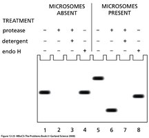
Q1. Explain the experimental results seen in lane 1-5. How would you account for the migration of proteins in lane 5, 6 and 8? Is the protein anchored in the membrane or is it translocated in the ER lumen? Explain your reasoning.
Background
Topic: Protein translocation across the endoplasmic reticulum (ER) membrane
This question tests your understanding of how proteins are translocated into the ER, and how their location and processing can be determined using protease protection, glycosylation, and signal peptide cleavage. The experiment uses microsomes (ER membrane vesicles), protease, detergent, and endoglycosidase H (endo H) to analyze protein processing and localization.
Key Terms and Concepts:
Microsomes: Vesicles derived from fragmented ER membranes.
Protease: Enzyme that digests proteins; only accessible proteins are degraded.
Detergent: Disrupts membranes, exposing all proteins to protease.
Endo H: Enzyme that removes certain types of N-linked oligosaccharides.
Glycosylation: Addition of sugar groups to proteins in the ER lumen.
Signal peptide cleavage: Removal of the signal sequence after translocation.
Step-by-Step Guidance
Examine the treatments for each lane (1-8) and note which conditions are present: microsomes, protease, detergent, endo H.
For lanes 1-5, compare the migration pattern of the protein bands. Consider how the presence or absence of microsomes and protease affects protein protection and processing.
Recall that proteins protected from protease are likely inside microsomes (ER lumen), while those degraded are exposed (cytosolic or membrane-bound).
Analyze the effect of detergent: it disrupts the membrane, exposing all proteins to protease, so bands may disappear or shift.
For lanes with endo H treatment, consider how glycosylation affects migration. Endo H removes certain sugars, causing a shift in band position.
For lanes 5, 6, and 8, focus on how the combination of treatments (microsomes, protease, detergent, endo H) influences the migration and what this reveals about protein localization and processing.
Try solving on your own before revealing the answer!
Final Answer:
Lanes 1-5: Lane 1 shows the protein synthesized without microsomes (cytosolic). Lane 2 shows the protein is degraded by protease (no protection). Lane 3 shows the protein is protected from protease when microsomes are present, indicating translocation into the ER lumen. Lane 4 shows that detergent disrupts the membrane, exposing the protein to protease and resulting in degradation. Lane 5 shows a shift in migration after endo H treatment, indicating glycosylation and subsequent removal of sugars.
Lanes 5, 6, and 8: The migration in lane 5 (endo H treated) is faster due to removal of oligosaccharides. Lane 6 (microsomes + protease + endo H) shows a protected, deglycosylated protein. Lane 8 (microsomes + detergent + endo H) shows deglycosylated protein exposed by detergent.
Protein localization: The protein is translocated into the ER lumen, as evidenced by protection from protease in the presence of microsomes and glycosylation. It is not anchored in the membrane, since it is fully protected and processed.
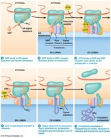
Q2. Describe the status of translation, co-translational import, and pore (open or closed) for each treatment: a) Wild-type SRP with GppNp, b) Mutant SRP with GTP, c) Mutant SRP with GppNp.
Background
Topic: Mechanism of co-translational protein import into the ER
This question tests your understanding of how the signal recognition particle (SRP), GTP hydrolysis, and ribosome-membrane interactions regulate protein import into the ER. The experiment uses wild-type and mutant SRP, and GTP analogs to dissect the process.
Key Terms and Concepts:
SRP: Signal recognition particle, binds signal sequence and pauses translation.
GTP/GDP: Guanine nucleotides, regulate SRP function.
GppNp: Non-hydrolyzable GTP analog, prevents GTP hydrolysis.
Co-translational import: Protein is imported into ER during translation.
Pore: Translocon channel in ER membrane, opens for import.
Step-by-Step Guidance
For each treatment, identify the SRP type and nucleotide present (GTP, GDP, GppNp).
Recall that wild-type SRP requires GTP hydrolysis to release the ribosome and open the pore for import.
Mutant SRP with high GDP affinity may not allow proper GTP binding or hydrolysis, affecting translation and import.
GppNp prevents GTP hydrolysis, so steps requiring hydrolysis may be blocked.
For each scenario, predict whether translation proceeds, whether co-translational import occurs, and whether the pore is open or closed, based on SRP and nucleotide status.
Try solving on your own before revealing the answer!
Final Answer:
a) Wild-type SRP with GppNp: Translation is paused, co-translational import does not occur, pore remains closed.
b) Mutant SRP with GTP: Translation proceeds, co-translational import does not occur, pore remains closed.
c) Mutant SRP with GppNp: Translation is paused, co-translational import does not occur, pore remains closed.
GTP hydrolysis is essential for SRP release and pore opening; blocking this step prevents import.
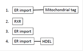
Q3. Predict the final destination of proteins encoded by genes 1, 2, 3, and 4, and which cellular compartments these proteins will go through before reaching their final destination.
Background
Topic: Protein targeting and sorting signals
This question tests your understanding of how signal sequences direct proteins to specific cellular compartments, and the pathway they follow through the endomembrane system.
Key Terms and Concepts:
ER import: Signal sequence directs protein to the endoplasmic reticulum.
Mitochondrial tag: Directs protein to mitochondria.
RXR: Retention signal for ER or Golgi.
HDEL: ER retention/retrieval signal.
Step-by-Step Guidance
For each protein, identify the signal sequences present and their order.
Recall that ER import signal directs proteins to the ER, where further sorting occurs based on additional tags.
For protein 1, consider whether ER import or mitochondrial tag dominates, and which compartment is reached first.
For protein 2, RXR signal may cause retention in ER or Golgi.
For protein 3, ER import signal alone directs to ER, with possible further transport.
For protein 4, ER import followed by HDEL signal results in ER retention or retrieval from Golgi.
Try solving on your own before revealing the answer!
Final Answer:
Protein 1: Initially imported into ER, but mitochondrial tag may redirect to mitochondria after ER entry.
Protein 2: RXR signal causes retention in ER or Golgi; protein does not proceed further.
Protein 3: Imported into ER, may be secreted or processed further depending on additional signals.
Protein 4: Imported into ER, HDEL signal ensures retention or retrieval to ER from Golgi.
Proteins follow the pathway dictated by their signal sequences, moving through ER, Golgi, and other compartments as specified.
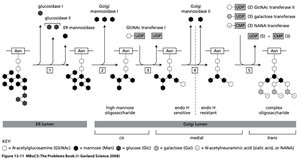
Q4. Arrange the mutants in order that corresponds to the steps in pathway for processing N-linked oligosaccharides. Assume each mutant is defective for a single enzyme only.
Background
Topic: N-linked glycosylation and oligosaccharide processing in the ER and Golgi
This question tests your understanding of the sequential steps in N-linked oligosaccharide processing, and how defects in specific enzymes affect the sugar composition of glycoproteins.
Key Terms and Concepts:
N-linked glycosylation: Attachment of oligosaccharides to asparagine residues in proteins.
Man: Mannose, GlcNAc: N-acetylglucosamine, Gal: Galactose, NANA: Sialic acid, Glc: Glucose.
Enzymes: Glycosidases and glycosyltransferases remove or add sugars in a specific order.
Step-by-Step Guidance
Examine the sugar composition for each mutant and compare to wild type.
Identify which sugars are missing or present in excess, indicating the step where processing is blocked.
Refer to the pathway diagram to match each mutant to a specific enzymatic step.
Arrange the mutants in order from earliest to latest step in the pathway, based on their sugar profiles.
Check for mutants with high mannose or glucose, indicating early defects, and those missing galactose or NANA, indicating later defects.
Try solving on your own before revealing the answer!
Final Answer:
The order of mutants corresponds to the pathway steps as follows (from earliest to latest defect):
Mutant D/H/G/C (high mannose, early ER processing defect)
Mutant E (intermediate processing, less GlcNAc)
Mutant A/I (defective GlcNAc transfer, no galactose or NANA)
Mutant B (defective galactose transfer)
Mutant F (defective NANA transfer)
Each mutant blocks a specific step, resulting in characteristic sugar profiles.
