 Back
Study Guide: The Macromolecules of the Cell – Protein Structure and Function
Study Guide - Smart Notes
 Tailored notes based on your materials, expanded with key definitions, examples, and context.
The Macromolecules of the Cell
Overview of Biological Macromolecules
Most biological macromolecules in cells are synthesized from a limited set of small molecules, which are assembled into polymers with diverse functions. - Key Point 1: Macromolecules include proteins, nucleic acids, polysaccharides, and lipids. - Key Point 2: These macromolecules are built from monomeric units such as amino acids, nucleotides, sugars, and fatty acids. - Example: Proteins are polymers of amino acids, while nucleic acids are polymers of nucleotides. 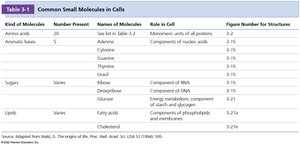
Protein Structure and Function
Classes of Proteins
Proteins serve a wide range of functions in cells, each class specialized for a particular role. - Enzymes: Catalyze biochemical reactions, increasing reaction rates. - Structural proteins: Provide physical support and shape to cells and tissues. - Motility proteins: Enable contraction and movement. - Regulatory proteins: Control and coordinate cellular functions. - Transport proteins: Move substances across cellular membranes. - Signaling proteins: Facilitate communication between cells. - Receptor proteins: Allow cells to respond to environmental stimuli. - Defensive proteins: Protect against disease. - Storage proteins: Serve as reservoirs of amino acids.
Amino Acids: The Monomers of Proteins
Proteins are synthesized from 20 standard amino acids, each with a unique side chain (R group) that determines its properties. - Key Point 1: All amino acids share a common structure: a central α-carbon, an amino group, a carboxyl group, and a variable R group. - Key Point 2: In biological systems, amino acids are almost exclusively found in the L-form. 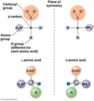
Chemical Structure and Classification of Amino Acids
Amino acids are classified based on the chemical nature of their side chains: nonpolar, polar uncharged, and polar charged. - Key Point 1: The side chain determines the amino acid's role in protein structure and function. - Key Point 2: Glycine is unique among nonpolar amino acids as it does not have separate L and D isomers due to its symmetric structure. 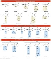
Abbreviations for Amino Acids
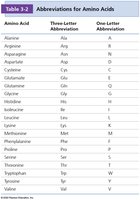
Amino acids are commonly referred to by their three-letter and one-letter abbreviations, which are essential for interpreting protein sequences.
Amino Acid | Three-Letter Abbreviation | One-Letter Abbreviation |
|---|---|---|
Alanine | Ala | A |
Arginine | Arg | R |
Asparagine | Asn | N |
Aspartic acid | Asp | D |
Cysteine | Cys | C |
Glutamine | Gln | Q |
Glutamic acid | Glu | E |
Glycine | Gly | G |
Histidine | His | H |
Isoleucine | Ile | I |
Leucine | Leu | L |
Lysine | Lys | K |
Methionine | Met | M |
Phenylalanine | Phe | F |
Proline | Pro | P |
Serine | Ser | S |
Threonine | Thr | T |
Tryptophan | Trp | W |
Tyrosine | Tyr | Y |
Valine | Val | V |
Peptide Bond Formation
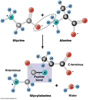
Amino acids are linked together by peptide bonds, formed through dehydration (condensation) reactions. - Key Point 1: The peptide bond is a covalent bond between the carboxyl group of one amino acid and the amino group of another. - Key Point 2: Peptide bond formation releases a molecule of water. 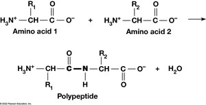
Monomeric and Multimeric Proteins
Proteins may consist of a single polypeptide (monomeric) or multiple polypeptides (multimeric). - Key Point 1: Multimeric proteins are classified by the number of polypeptide subunits: dimers (2), trimers (3), tetramers (4), etc. - Example: Hemoglobin is a tetramer composed of two α and two β subunits. 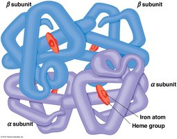
Bonds and Interactions in Protein Folding and Stability
Protein folding and stability depend on both covalent and noncovalent interactions. - Covalent bonds: Peptide bonds and disulfide bonds. - Non-covalent interactions: Hydrogen bonds, ionic bonds, van der Waals forces, and hydrophobic interactions. - Key Point 2: These interactions involve the carboxyl, amino, and R groups of amino acid residues. 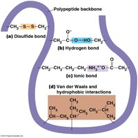
Levels of Protein Structure
Overview of Structural Levels
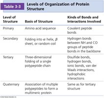
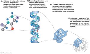
Protein structure is organized into four hierarchical levels: primary, secondary, tertiary, and quaternary.
Level of Structure | Basis of Structure | Kinds of Bonds and Interactions Involved |
|---|---|---|
Primary | Amino acid sequence | Covalent peptide bonds |
Secondary | Folding into α helix, β sheet, or random coil | Hydrogen bonds between NH and CO groups of peptide bonds in the backbone |
Tertiary | Three-dimensional folding of a single polypeptide chain | Disulfide bonds, hydrogen bonds, ionic bonds, van der Waals interactions, hydrophobic interactions |
Quaternary | Association of multiple polypeptides to form a multimeric protein | Same as for tertiary structure |
Primary Structure
The primary structure is the linear sequence of amino acids in a polypeptide, written from the N-terminus to the C-terminus. - Key Point 1: The sequence is genetically determined by the order of nucleotides in mRNA. - Key Point 2: The order and identity of amino acids direct the formation of higher-order structures. 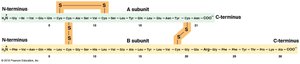
Secondary Structure
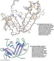
Secondary structure refers to local regions of folding stabilized by hydrogen bonding between backbone NH and CO groups. - Key Point 1: The two major patterns are the α helix and the β sheet. - Key Point 2: Certain amino acids favor specific secondary structures; proline disrupts α helices due to its inability to form hydrogen bonds. 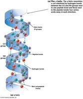
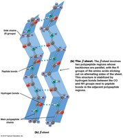
Common Secondary Motifs
Motifs are supersecondary structures composed of a few secondary structure elements, such as β-α-β motifs, hairpin loops, and helix-turn-helix motifs. 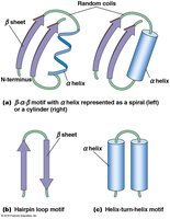
Tertiary Structure
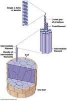
Tertiary structure is the overall three-dimensional shape of a single polypeptide, determined by interactions among R groups. - Key Point 1: Hydrophobic residues avoid water, hydrophilic residues interact with water, and charged residues attract or repel each other. - Key Point 2: Fibrous proteins (e.g., keratin, fibroin) have extensive regions of secondary structure, while globular proteins are compact and folded. 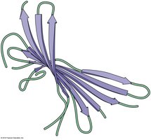
Protein Domains
Domains are distinct structural and functional units within a protein. - Key Point 1: Proteins with similar functions often share common domains. - Key Point 2: Multifunctional proteins may have separate domains for each function. 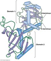
Quaternary Structure
Quaternary structure describes the association of multiple polypeptide chains to form a multimeric protein. - Key Point 1: The same types of bonds and interactions as in tertiary structure maintain quaternary structure. - Example: Hemoglobin's quaternary structure consists of two α and two β subunits.
Protein Folding and Disease
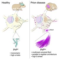
Misfolded proteins can lead to disease, such as prion diseases, which involve abnormal protein aggregation. 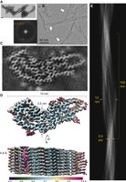

Summary Table: Levels of Protein Structure
Level | Basis | Bonds/Interactions |
|---|---|---|
Primary | Amino acid sequence | Peptide bonds |
Secondary | α helix, β sheet | Hydrogen bonds |
Tertiary | 3D folding | Disulfide, hydrogen, ionic, van der Waals, hydrophobic |
Quaternary | Subunit assembly | Same as tertiary |
Additional info: AlphaFold is a modern AI system for predicting protein tertiary structure, accelerating research in cell biology.
