 Back
Back22:The Cytoskeleton II: Myosins, Muscle Contraction, and the Sliding Filament Mechanism
Study Guide - Smart Notes
 Tailored notes based on your materials, expanded with key definitions, examples, and context.
The Cytoskeleton II: Myosins and Muscle Contraction
Myosins: Actin-Binding Motor Proteins
Myosins are a superfamily of actin-dependent motor proteins that play essential roles in cellular movement and muscle contraction. They convert chemical energy from ATP hydrolysis into mechanical work, moving along actin filaments.
All actin-dependent motor proteins belong to the myosin family.
ATP hydrolysis by myosins provides the energy for movement along actin filaments.
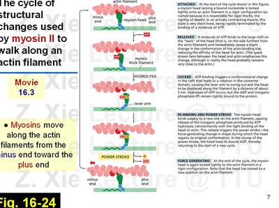
Myosins typically move from the minus end toward the plus end of actin filaments.
Structure of Myosin II
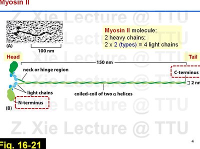
Myosin II is the primary myosin involved in muscle contraction. It is a dimeric protein composed of two heavy chains and four light chains, forming a coiled-coil structure.
Heavy chains: Form the long coiled-coil tail and globular head domains.
Light chains: Bind near the neck region and regulate myosin activity.
Head domain: Contains the actin-binding and ATPase activity sites.
Tail domain: Facilitates dimerization and filament assembly.
Myosin II Bipolar Thick Filament in Muscle
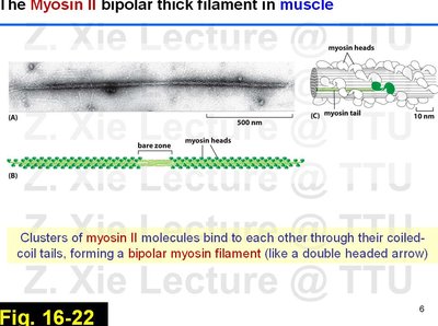
In muscle cells, myosin II molecules assemble into bipolar thick filaments, which are essential for muscle contraction. These filaments are formed by the association of myosin tails, creating a structure with heads at both ends.
Bipolar filament: Myosin heads project outward from both ends, allowing interaction with actin filaments.
Coiled-coil tails: Central region where tails overlap, forming the filament's core.
The Myosin II Cross-Bridge Cycle
Muscle contraction is driven by the cyclic interaction of myosin heads with actin filaments, known as the cross-bridge cycle. This process is powered by ATP hydrolysis and involves several conformational changes in the myosin head.
Attached: Myosin head is tightly bound to actin (rigor state).
Released: ATP binds to myosin, causing it to release from actin.
Cocked: ATP hydrolysis causes the myosin head to move to a "cocked" position.
Force-generating (Power Stroke): Myosin binds to actin, releases inorganic phosphate, and the head pivots, pulling the actin filament.
ADP release: Myosin remains attached until a new ATP binds, restarting the cycle.
Organization of Sarcomeres and Myofibrils
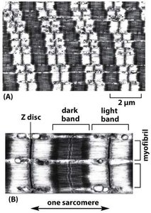
Sarcomere Structure
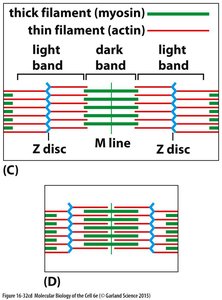
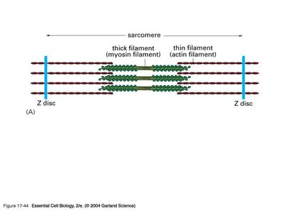
The sarcomere is the fundamental contractile unit of striated muscle, composed of highly organized arrays of actin (thin) and myosin (thick) filaments. The regular arrangement of these filaments gives muscle its striated appearance.
Z disc: Defines the boundaries of each sarcomere; attachment site for the plus ends of actin filaments.
M line: Center of the sarcomere; site of myosin filament cross-linking.
Light band (I band): Contains only thin filaments (actin).
Dark band (A band): Contains thick filaments (myosin), with overlapping thin filaments.
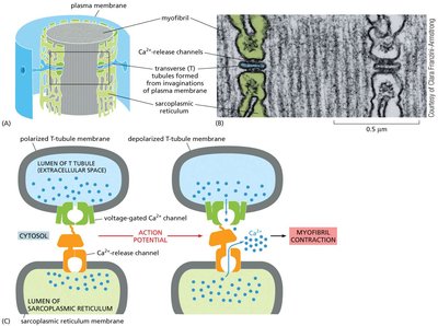
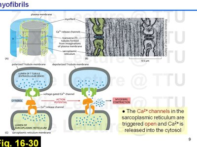
Excitation-Contraction Coupling
Muscle contraction is initiated by a nerve impulse that triggers the release of calcium ions (Ca2+) from the sarcoplasmic reticulum into the cytosol. This process is known as excitation-contraction coupling.
T tubules: Invaginations of the plasma membrane that transmit action potentials into the muscle fiber.
Sarcoplasmic reticulum: Specialized endoplasmic reticulum that stores Ca2+.
Ca2+ release: Voltage-gated channels open in response to depolarization, releasing Ca2+ into the cytosol.
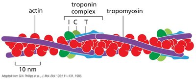
Regulation of Muscle Contraction: Role of Troponin and Tropomyosin
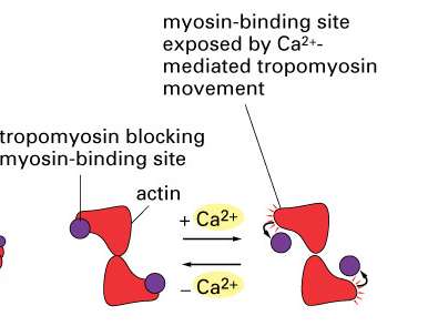
Muscle contraction is regulated by the interaction of Ca2+ with the proteins troponin and tropomyosin, which control the accessibility of myosin-binding sites on actin filaments.
Tropomyosin: Binds along actin filaments, blocking myosin-binding sites in the absence of Ca2+.
Troponin: A complex of three proteins, including a Ca2+-sensitive subunit. When Ca2+ binds, troponin induces a conformational change in tropomyosin, exposing the myosin-binding sites on actin.
Muscle Contraction: The Sliding Filament Mechanism
Muscle contraction occurs through the sliding filament mechanism, where actin and myosin filaments slide past each other, shortening the sarcomere without changing the length of the individual filaments.
Myosin heads walk toward the plus end of actin filaments, pulling the thin filaments toward the center of the sarcomere.
Simultaneous shortening of all sarcomeres in a muscle fiber results in overall muscle contraction.
Summary Table: Key Proteins in Muscle Contraction
Protein | Function |
|---|---|
Myosin II | Motor protein that interacts with actin to generate force for contraction |
Actin | Forms thin filaments; provides track for myosin movement |
Tropomyosin | Blocks myosin-binding sites on actin in resting muscle |
Troponin | Ca2+-sensitive complex that regulates tropomyosin position |
Sarcoplasmic Reticulum | Stores and releases Ca2+ to trigger contraction |
Additional info: The sliding filament model is fundamental to understanding muscle physiology and is a classic example of how molecular motors convert chemical energy into mechanical work in cells.
