 Back
BackThe Cytoskeleton: Structure, Function, and Clinical Correlations
Study Guide - Smart Notes
 Tailored notes based on your materials, expanded with key definitions, examples, and context.
The Cytoskeleton
Introduction and Overview
The cytoskeleton is a dynamic network of protein filaments that provides structural support, facilitates cellular movement, and organizes intracellular transport. It is essential for maintaining cell shape, enabling cell division, and supporting various cellular functions. The cytoskeleton is composed of three main types of protein filaments: microfilaments (actin filaments), intermediate filaments, and microtubules, each with distinct structures and functions.
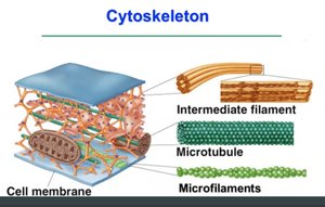
Microfilaments (Actin Filaments): Thin, flexible fibers involved in cell movement and shape.
Intermediate Filaments: Rope-like fibers providing mechanical strength and resistance to stress.
Microtubules: Hollow tubes that organize the cell’s interior and serve as tracks for intracellular transport.
Microfilaments (Actin Filaments)
Structure and Assembly
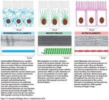
Microfilaments are composed of G-actin (globular actin) monomers that polymerize to form F-actin (filamentous actin) polymers. These filaments are approximately 7 nm in diameter and exhibit polarity, with a fast-growing (+) barbed end and a slower-growing (–) pointed end. The assembly and disassembly of actin filaments are ATP-dependent and highly dynamic, a process known as treadmilling.
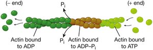
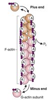
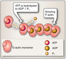
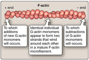
Treadmilling: Addition of ATP-bound actin at the (+) end and dissociation of ADP-bound actin at the (–) end.
Regulation: Controlled by actin-binding proteins such as profilin (promotes assembly), cofilin (promotes disassembly), and capping proteins.
Functions of Microfilaments
Actin filaments are crucial for various cellular processes, including:
Cell motility (e.g., migration, chemotaxis)
Muscle contraction (interaction with myosin)
Cytokinesis (formation of the contractile ring during cell division)
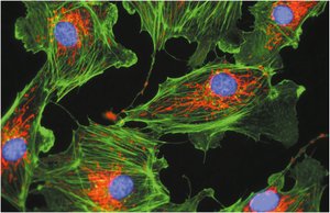
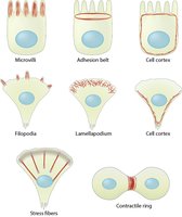
Maintenance of cell shape and formation of cellular protrusions (e.g., microvilli, lamellipodia)
Wound healing
Actin Dynamics and Regulation
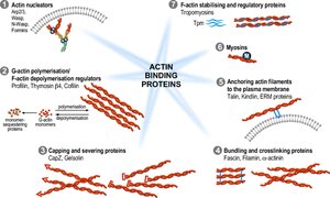
The dynamic behavior of actin filaments is regulated by a variety of actin-binding proteins:
Nucleation factors: Arp2/3 complex, formins, and spire proteins initiate new filament formation.
Polymerization factors: Profilin, cofilin, and thymosin regulate monomer addition and removal.
Stabilizing proteins: Tropomyosins stabilize F-actin.
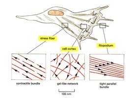
Crosslinking proteins: α-actinin, filamin, and fascin organize filaments into networks or bundles.
Severing proteins: Gelsolin and CapZ fragment filaments to remodel the cytoskeleton.
Specialized Actin Structures and Functions
Cell Shape Diversity: Actin filaments enable cells to adopt various shapes and form specialized structures such as microvilli, filopodia, and stress fibers.
Mechanical Strength: Cross-linked actin networks increase cellular mechanical strength.
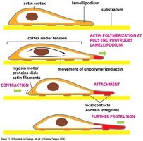
Cell Migration: Actin polymerization at the leading edge pushes the plasma membrane forward, while contraction at the rear moves the cell body.
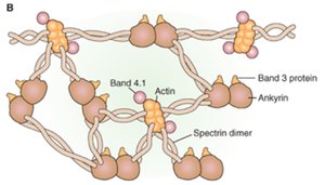
Actin in Red Blood Cells: Spectrin Meshwork
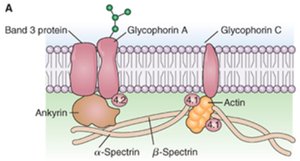
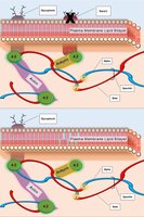
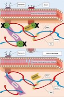
In erythrocytes, a spectrin-actin meshwork forms the cell cortex, providing flexibility and durability. Spectrin and actin are linked to the plasma membrane via ankyrin and other proteins, maintaining cell shape and membrane integrity.
Clinical Correlation: Hereditary Spherocytosis
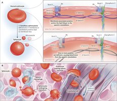
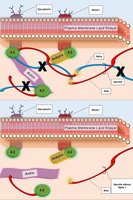
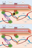
Hereditary spherocytosis is a genetic disorder caused by mutations in genes encoding spectrin, ankyrin, band 3, or protein 4.2. These mutations disrupt the cytoskeletal network, leading to red blood cell membrane instability, hemolytic anemia, jaundice, and splenomegaly.
Clinical Correlation: Muscular Dystrophy
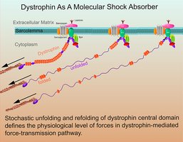
Muscular dystrophies are a group of genetic disorders characterized by muscle membrane instability, often due to defects in the dystrophin protein complex. Dystrophin links the actin cytoskeleton to the extracellular matrix, and its absence leads to progressive muscle weakness.
Intermediate Filaments
Structure and Types
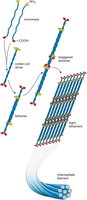
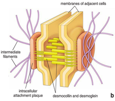
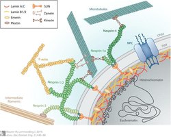
Intermediate filaments are rope-like, non-polar protein fibers approximately 10 nm in diameter. They provide mechanical stability and resistance to stress, especially in tissues subject to mechanical forces. Intermediate filaments are found at cell junctions (desmosomes) and in the nuclear lamina.
Functions of Intermediate Filaments
Mechanical support and stress resistance
Nuclear organization (nuclear lamina)
Cell-cell adhesion (desmosomes)
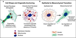
Tissue integrity and organelle anchoring
Types and Distribution
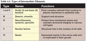
Type I/II: Keratins (epithelial cells)
Type III: Vimentin (mesenchymal cells)
Type IV: Neurofilaments (neurons)
Type V: Nuclear lamins (all cells)
Lamins and Nuclear Structure
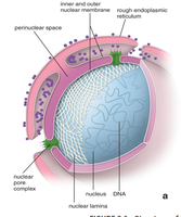
Lamins are intermediate filaments located beneath the nuclear envelope, anchoring chromatin and organizing the nucleus. They are essential for nuclear envelope reassembly after mitosis and for DNA replication.
Clinical Correlations of Intermediate Filaments
Epidermolysis bullosa simplex: Mutations in keratin genes cause skin blistering and nail deformities.
Alexander disease: Mutation in GFAP (astrocyte intermediate filament) leads to leukodystrophy and neurological symptoms.
Progeria syndrome: Mutation in the LMNA gene (lamin A) causes premature aging due to nuclear envelope defects.
Microtubules
Structure and Organization
Microtubules are hollow tubes composed of α- and β-tubulin dimers, with a diameter of 25 nm. They are polarized structures with dynamic instability, meaning they rapidly grow and shrink. Microtubules are organized by microtubule organizing centers (MTOCs), such as the centrosome.
Dynamic instability: GTP-bound tubulin adds to the plus end, while GTP hydrolysis leads to rapid depolymerization if the GTP cap is lost.
Drugs: Colchicine prevents assembly; taxol prevents disassembly.
Functions of Microtubules
Intracellular transport (vesicles, organelles)
Cell division (mitotic spindle formation)
Cell shape maintenance
Ciliary and flagellar movement
Axonal transport in neurons
Motor Proteins and Transport
Microtubule-associated motor proteins include kinesins (anterograde transport) and dyneins (retrograde transport). These proteins use ATP hydrolysis to move cargo along microtubules, including vesicles, organelles, and proteins.
Cilia and Flagella
Cilia and flagella are microtubule-based structures with a characteristic 9+2 arrangement. They are involved in mucus clearance, left-right patterning, sensory reception, and cell signaling.
Clinical Correlations: Ciliopathies
Primary ciliary dyskinesia: Autosomal recessive disorder causing abnormal ciliary movement, leading to chronic respiratory disease, sinusitis, otitis media, and infertility.
Polycystic kidney disease: Mutations in PKD1/PKD2 disrupt ciliary function, causing cyst formation, hypertension, and kidney failure.
