 Back
BackCh. 12: The Endomembrane System and Protein Sorting: Structure, Function, and Trafficking
Study Guide - Smart Notes
 Tailored notes based on your materials, expanded with key definitions, examples, and context.
The Endomembrane System
Overview of the Endomembrane System
The endomembrane system is a network of membranous organelles in eukaryotic cells that coordinates the synthesis, processing, and transport of proteins and lipids. It includes the endoplasmic reticulum (ER), Golgi apparatus, endosomes, lysosomes, nuclear envelope, plasma membrane, and peroxisomes. - Key Components: ER, Golgi apparatus, endosomes, lysosomes, nuclear envelope, plasma membrane, peroxisomes - Function: Integration of biosynthetic, sorting, and trafficking pathways for proteins and lipids 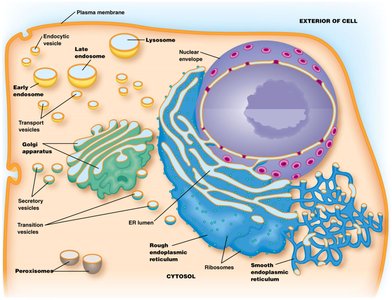
Endoplasmic Reticulum (ER)
Structure and Types of ER
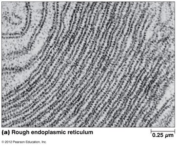
The ER is a continuous membrane system divided into rough and smooth regions, each with distinct functions. - Rough ER: Studded with ribosomes; involved in protein synthesis and processing - Smooth ER: Lacks ribosomes; involved in lipid synthesis, detoxification, and calcium storage 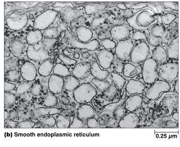
Functions of the ER
The ER is central to the biosynthesis and processing of proteins and lipids. - Protein Biosynthesis: Synthesis, folding, and assembly of polypeptides; removal of misfolded proteins - Lipid Biosynthesis: Primary source of membrane lipids in eukaryotic cells - Carbohydrate Metabolism: Initial steps of glycoprotein formation - Detoxification: Drug metabolism in smooth ER - Calcium Storage: Regulation of intracellular calcium levels - Steroid Biosynthesis: Synthesis of steroid hormones in smooth ER
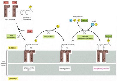
ER Membrane Structure and Lipid Synthesis
The ER membrane consists of cisternae (membrane-bound sacs) and a lumen (internal space). Fatty acids are synthesized in the cytoplasm and incorporated into the ER membrane. Flippases transfer lipids from the cytosolic to the lumenal side of the bilayer, ensuring growth of both halves of the membrane. 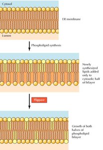
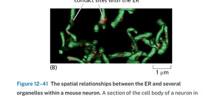
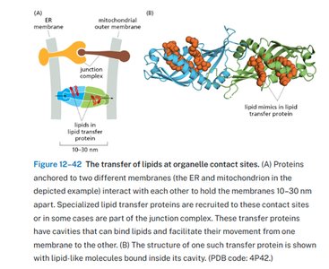
Membrane Contact Sites and Lipid Transfer
The ER forms contact sites with other organelles, facilitating selective lipid transfer via lipid transfer proteins. These proteins move specific phospholipids to mitochondria, chloroplasts, and peroxisomes. 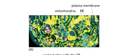
Composition of ER and Plasma Membranes
The ER and plasma membranes differ in their lipid and protein composition, reflecting their specialized functions.
Membrane Components | ER Membrane | Plasma Membrane |
|---|---|---|
Carbohydrate (% by weight) | 10 | 10 |
Protein (% by weight) | 62 | 54 |
Total lipid (% by weight) | 27 | 36 |
Phosphatidylcholine (% of total lipids) | 40 | 24 |
Phosphatidylethanolamine (% of total lipids) | 17 | 7 |
Phosphatidylserine (% of total lipids) | 5 | 4 |
Cholesterol (% of total lipids) | 6 | 17 |
Sphingomyelin (% of total lipids) | 5 | 19 |
Glycolipids (% of total lipids) | trace | 7 |
Other lipids (% of total lipids) | 27 | 22 |
The Golgi Complex
Structure and Organization
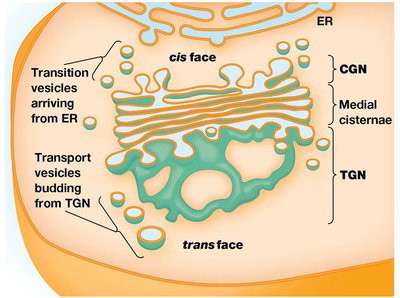
The Golgi complex is a series of flattened, membrane-bounded cisternae forming a stack. It is divided into the cis-Golgi network (CGN), medial cisternae, and trans-Golgi network (TGN). - CGN: Receives vesicles from the ER - Medial cisternae: Central region for processing - TGN: Sorts and dispatches vesicles to their destinations 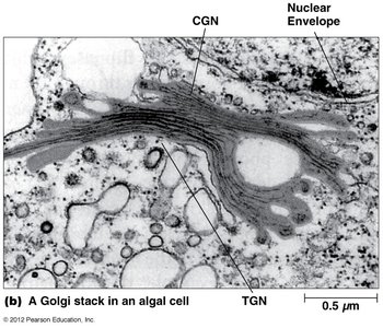
Models of Golgi Trafficking
Two models explain the flow of lipids and proteins through the Golgi apparatus: - Stationary Cisternae Model: Golgi cisternae are stable; shuttle vesicles mediate transport between cisternae - Cisternal Maturation Model: Cisternae are transient; they mature from CGN to TGN, with enzymes returned to earlier compartments 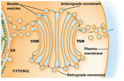
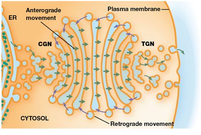
Anterograde and Retrograde Transport
- Anterograde Transport: Movement of material toward the plasma membrane - Retrograde Transport: Flow of vesicles from Golgi cisternae back to the ER 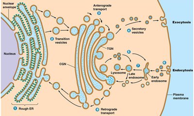
Protein Processing in the ER and Golgi
Protein Folding and Quality Control
Proteins fold and assemble in the ER lumen, assisted by molecular chaperones (e.g., BiP). Misfolded proteins trigger the unfolded protein response (UPR), which shuts down protein production and activates chaperone genes. 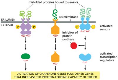
Glycosylation
Glycosylation is the addition of carbohydrate groups to proteins, critical for their function and sorting. - N-linked glycosylation: Addition of oligosaccharide to the nitrogen atom of asparagine residues - O-linked glycosylation: Addition of oligosaccharide to the oxygen atom of serine, threonine, or tyrosine residues - Compartmentalization: Initial steps occur in the ER; further modifications occur in the Golgi 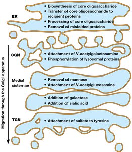
Trafficking Through the Endomembrane System
Vesicular Transport and Sorting
Proteins and lipids are transported between organelles via vesicular tubular clusters and transport vesicles. - ER-Golgi Intermediate Complex: Mediates transport from ER to Golgi - Retention and Retrieval Tags: ER-specific proteins contain retention (RXR) and retrieval (KDEL, KKXX, HDEL) tags to maintain localization - Golgi Complex Proteins: Retention/retrieval tags and hydrophobic domain length determine localization
Lysosomes and Cellular Digestion
Lysosomes are organelles containing digestive enzymes for degradation of external materials (phagocytosis, receptor-mediated endocytosis) and cellular structures (autophagy). Lysosomes develop from endosomes, with enzymes delivered from the TGN.
Exocytosis and Endocytosis
- Exocytosis: Release of intracellular molecules outside the cell - Endocytosis: Import of extracellular molecules by vesicle formation from the plasma membrane
Coated Vesicles and SNARE Proteins
Coated vesicles (clathrin, COPI, COPII) mediate vesicular traffic. ARF and Sar GTPases promote vesicle formation. SNARE proteins (v-SNAREs and t-SNAREs) mediate fusion between vesicles and target membranes, with Rab GTPases ensuring specificity. Example: Clathrin-coated vesicles transport lysosomal enzymes; SNAREs ensure fusion with lysosomes. Additional info: The notes include inferred details on the mechanisms of protein sorting and vesicular trafficking for completeness.
