 Back
BackThe Endomembrane System: Structure, Function, and Protein Trafficking
Study Guide - Smart Notes
 Tailored notes based on your materials, expanded with key definitions, examples, and context.
The Endomembrane System
Overview and Importance
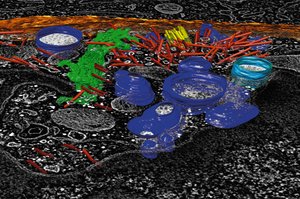
The endomembrane system is a collection of membranous organelles in eukaryotic cells that compartmentalize cellular functions and facilitate the regulated movement (trafficking) of lipids and proteins. Understanding this system is essential for appreciating the complexity and specialization of eukaryotic cells.
Trafficking refers to the movement of lipids and proteins between organelles, which must be tightly regulated.
Compartmentalization allows for specialized functions within organelles.
Components of the Endomembrane System
The endomembrane system includes several interconnected organelles and vesicles, each with distinct roles in protein and lipid processing, sorting, and degradation.
Endoplasmic Reticulum (ER): Site of protein and lipid synthesis.
Golgi Complex: Processes, sorts, and packages proteins and lipids.
Endosomes: Sort material brought into the cell.
Lysosomes: Digest ingested material and unneeded cellular components.
Vesicles connect these organelles, facilitating transport.
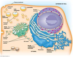
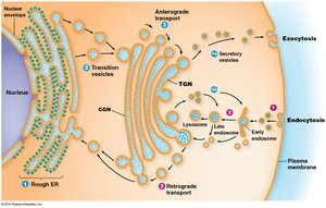
The Endoplasmic Reticulum (ER)
Structure and Function
The ER is a continuous network of flattened sacs, tubules, and vesicles. The membrane-bound sacs are called ER cisternae, and the space inside them is the ER lumen. The ER is involved in biosynthesis of proteins and lipids.
Proteins synthesized for incorporation into membranes, organelles, or export.
Lipid synthesis occurs in the ER.
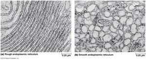
Rough vs. Smooth ER
The ER is divided into two types based on structure and function:
Rough ER: Studded with ribosomes; large, flattened sheets; involved in protein synthesis.
Smooth ER: No ribosomes; smooth, tubular structures; involved in lipid metabolism and detoxification.
The lumenal spaces of rough and smooth ER are continuous.
Variation in ER Types
The relative amounts of rough and smooth ER vary by cell type:
Cells synthesizing secretory proteins have prominent rough ER.
Cells producing steroid hormones have extensive smooth ER.
Functions of Rough ER
Rough ER is central to protein biosynthesis and processing:
Ribosomes synthesize membrane-bound and soluble proteins for the endomembrane system.
Proteins are inserted cotranslationally through a pore complex.
Initial steps of glycoprotein formation, polypeptide folding, removal of misfolded proteins, and assembly of multimeric proteins occur here.
Functions of Smooth ER
Smooth ER is involved in various metabolic reactions:
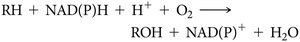
Drug Detoxification: Hydroxylation increases solubility of hydrophobic drugs, catalyzed by cytochrome P-450 monooxygenases.
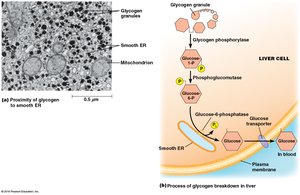
Carbohydrate Metabolism: Breakdown of glycogen; contains glucose-6-phosphatase.
Calcium Storage: Sarcoplasmic reticulum in muscle cells stores calcium ions.
Steroid Biosynthesis: Cholesterol and steroid hormone synthesis; HMG-CoA reductase is a key enzyme.
Membrane Biosynthesis in the ER
Source of Membrane Lipids
The ER is the primary source of membrane lipids in eukaryotic cells, with some exceptions (mitochondria, peroxisomes, chloroplasts).
Fatty acids for phospholipids are synthesized in the cytoplasm and incorporated into the ER membrane.
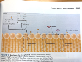
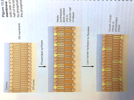
Phospholipid Translocation
Phospholipids are transferred to the lumenal side of the bilayer by phospholipid translocators (flippases).
Ensures even distribution of lipids across the bilayer.
The Golgi Complex
Structure and Function
The Golgi complex is functionally and physically linked to the ER. It processes, sorts, and packages glycoproteins and membrane lipids for transport.
Consists of a series of flattened membrane-bounded cisternae (Golgi stack).
Secretory cells may have hundreds or thousands of stacks.
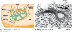
Cis and Trans Faces
The Golgi stack has two faces:
Cis face: Oriented toward the ER; cis-Golgi network (CGN).
Trans face: Oriented away from the ER; trans-Golgi network (TGN).
Anterograde and Retrograde Transport
Material moves through the Golgi via two main transport mechanisms:
Anterograde transport: Movement toward the plasma membrane (exocytosis).
Retrograde transport: Flow of vesicles from Golgi back to the ER, balancing membrane flow and supplying materials for new vesicles.
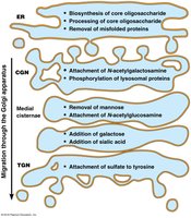
Protein Processing in the ER and Golgi
Protein Folding and Quality Control
Proteins fold and assemble in the ER lumen, assisted by molecular chaperones (e.g., BiP, protein disulfide isomerase). Quality control mechanisms detect and remove misfolded proteins.
Unfolded Protein Response (UPR): Detects misfolded proteins.
ER-associated degradation (ERAD): Exports misfolded proteins to the cytosol for degradation by proteasomes.
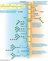
Protein Glycosylation
Glycosylation is the addition of carbohydrate side chains to proteins, forming glycoproteins. This process is coordinated between the ER and Golgi.
Initial steps of N-glycosylation occur on the cytosolic surface of the ER membrane.
Later steps occur in the ER lumen and are completed in the Golgi.
Protein Trafficking and Sorting
Protein Tags and Sorting Pathways
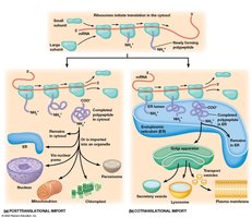
Proteins synthesized in the rough ER are directed to specific locations by protein tags (amino acid sequences, hydrophobic domains, oligosaccharide side chains). Two main pathways exist for sorting polypeptides:
Posttranslational import: Polypeptides destined for cytosol, mitochondria, chloroplast, peroxisome, or nucleus are synthesized by free ribosomes and imported after translation.
Cotranslational import: Polypeptides destined for the endomembrane system or export are synthesized by ribosomes attached to the ER membrane and inserted during synthesis.
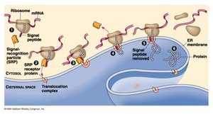
Signal Hypothesis and Cotranslational Import
The signal hypothesis states that polypeptides destined for the ER have an N-terminal ER signal sequence, which directs the ribosome-mRNA-polypeptide complex to the rough ER. The signal recognition particle (SRP) mediates this process.
SRP binds to the signal sequence and the ER membrane, blocking translation until the ribosome is docked.
The translocon complex facilitates transfer of the polypeptide into the ER lumen.
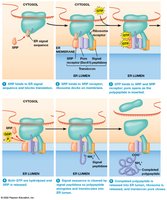
Steps of Cotranslational Import
Polypeptide synthesis begins; SRP binds signal sequence and halts translation.
SRP docks ribosome to translocon; GTP binding releases SRP and resumes translation.
Polypeptide enters ER lumen; signal peptidase removes signal peptide.
Polypeptide is released into ER lumen; ribosome detaches and dissociates.
Summary Table: Functions of the Endomembrane System Components
Organelle | Main Function |
|---|---|
Rough ER | Protein synthesis, folding, glycosylation, quality control |
Smooth ER | Lipid synthesis, drug detoxification, carbohydrate metabolism, calcium storage, steroid biosynthesis |
Golgi Complex | Processing, sorting, packaging of proteins and lipids |
Endosomes | Sorting of internalized material |
Lysosomes | Digestion of ingested material and cellular debris |
Additional info: The notes above expand on brief points with academic context, definitions, and examples to ensure completeness and clarity for cell-biology students.
