 Back
The Endomembrane System: Structure, Function, and Protein Processing
Study Guide - Smart Notes
 Tailored notes based on your materials, expanded with key definitions, examples, and context.
The Endomembrane System
Overview of the Endomembrane System
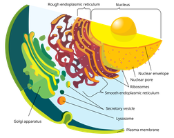
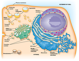
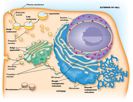
The endomembrane system is a collection of membranous organelles within eukaryotic cells that work together to modify, package, and transport proteins and lipids. Key components include the endoplasmic reticulum (ER), Golgi apparatus, endosomes, and lysosomes. These organelles are interconnected by vesicular transport and play essential roles in cellular homeostasis, secretion, and degradation.
Endoplasmic Reticulum (ER): Site of protein and lipid synthesis.
Golgi Apparatus: Modifies, sorts, and packages proteins and lipids for secretion or delivery to other organelles.
Endosomes: Involved in sorting and trafficking of internalized material.
Lysosomes: Responsible for degradation of cellular waste.
Endoplasmic Reticulum (ER)
Types and Structure of ER
The ER is a network of membranous tubules and sacs called cisternae. It is divided into two types based on structure and function:
Rough ER: Studded with ribosomes; involved in protein synthesis and processing.
Smooth ER: Lacks ribosomes; involved in lipid synthesis, detoxification, and calcium storage.
The lumen is the internal space of the ER where many reactions occur.
Rough Endoplasmic Reticulum: Functions
The rough ER is essential for the synthesis, folding, and trafficking of proteins destined for secretion, membrane insertion, or organelle targeting. It also detects improperly folded proteins and initiates quality control mechanisms.
Protein biosynthesis: Ribosomes on the rough ER synthesize polypeptides.
Protein processing: Folding and modification of proteins.
Protein trafficking: Transport of proteins to the Golgi apparatus.
Quality control: Detection and removal of misfolded proteins.
Protein Import into the ER
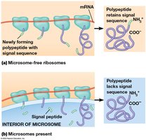
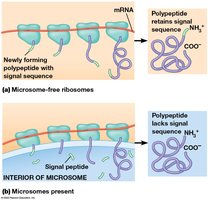
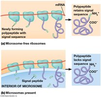
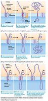
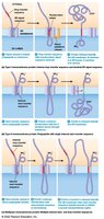
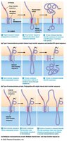
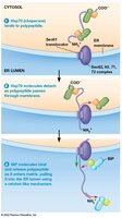
Proteins are imported into the ER either during translation (cotranslational import) or after translation (post-translational import). Cotranslational import involves the ER signal sequence, signal recognition particle (SRP), and the translocon complex.
Cotranslational import: Polypeptides are synthesized directly into the ER.
ER signal sequence: Directs ribosome to the ER membrane.
Signal recognition particle (SRP): Binds to the signal sequence and guides the ribosome to the ER.
Translocon: Protein channel in the ER membrane for polypeptide entry.
Post-Translational Import
Some proteins are imported into the ER after translation, assisted by chaperone proteins such as HSP70 and BiP. These chaperones bind to hydrophobic regions of the polypeptide, preventing aggregation and facilitating proper folding.
HSP70 chaperone: Prevents premature folding and aggregation.
BiP: ER-resident chaperone, member of the Hsp70 family.
Quality Control in the Rough ER
The ER employs several mechanisms to ensure only properly folded proteins proceed to the Golgi. Misfolded proteins are targeted for degradation via the unfolded protein response (UPR) and ER-associated degradation (ERAD), which involves proteasomes.
Unfolded Protein Response (UPR): Cellular response to accumulation of misfolded proteins.
ER-Associated Degradation (ERAD): Misfolded proteins are retrotranslocated to the cytosol and degraded by proteasomes.
Proteasomes: Multi-protein complexes that degrade unwanted proteins.
It is crucial for cells to remove improperly folded proteins to prevent cellular stress and disease.
Smooth Endoplasmic Reticulum: Functions
The smooth ER is involved in a variety of metabolic processes, including detoxification, carbohydrate metabolism, calcium storage, and steroid synthesis.
Detoxification: Hydroxylation reactions catalyzed by cytochrome P-450 enzymes, especially in the liver.
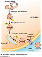
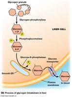
Carbohydrate metabolism: Breakdown of glycogen to glucose, important in liver cells.
Calcium storage: Regulates calcium ion concentration, crucial for muscle contraction.
Steroid synthesis: Produces steroid hormones in adrenal glands, testes, ovaries, and liver.
Enzymes Involved in Carbohydrate Metabolism
Several enzymes catalyze the breakdown of glycogen in the liver:
Phosphoglucomutase: Converts glucose-1-phosphate to glucose-6-phosphate.
Glycogen phosphorylase: Releases glucose-1-phosphate from glycogen.
Glucose-6-phosphatase: Converts glucose-6-phosphate to free glucose.
These reactions are essential for maintaining blood glucose levels.
Membrane Biosynthesis
The ER is the primary site for the synthesis of phospholipids and cholesterol, which are essential components of cellular membranes. Flippases are enzymes that help distribute lipids between the two leaflets of the bilayer.
Phospholipids: Major structural component of membranes.
Cholesterol: Modulates membrane fluidity.
Flippases: Catalyze the movement of lipids across the bilayer.
Golgi Apparatus
Structure and Function
The Golgi apparatus is composed of stacked, flattened membrane sacs called cisternae. It is responsible for modifying, sorting, and packaging proteins and lipids received from the ER.
Cis Golgi Network: Entry face, receives vesicles from ER.
Trans Golgi Network: Exit face, sorts and dispatches vesicles to their destinations.
Medial Cisternae: Intermediate processing compartments.
Models of Transport Through the Golgi
Several models explain how materials move through the Golgi:
Stationary Model: Cisternae remain fixed; vesicles transport materials between them.
Cisternal Maturation Model: Cisternae themselves mature and move forward.
Diffusion Model: Materials diffuse through the Golgi.
Kiss-and-Run Model: Vesicles transiently fuse with cisternae.
Transport Directions in the Golgi
Material moves in both anterograde (forward, ER to Golgi to plasma membrane) and retrograde (backward, Golgi to ER) directions. This bidirectional flow is necessary for recycling membrane components and maintaining organelle function.
Anterograde transport: Moves newly synthesized proteins and lipids forward.
Retrograde transport: Returns mis-sorted or recycled materials to the ER.
Protein Processing: Glycosylation
Glycosylation in ER and Golgi
Glycosylation is the addition of carbohydrate groups to proteins, which occurs in both the ER and Golgi. There are two main types:
N-linked glycosylation: Attachment to nitrogen atom on asparagine residue.
O-linked glycosylation: Attachment to oxygen atom on serine or threonine residue.
Functions of Glycosylation
Protein stability: Protects proteins from degradation.
Transport signal: Directs proteins to correct cellular locations.
Glycocalyx: Serves as markers and receptors on cell surface.
Glycosylation in the ER
In the ER, a core oligosaccharide is assembled and transferred to the protein. The process involves dolichol phosphate as a lipid carrier and sequential addition of sugars:
2 N-acetylglucosamine
9 mannose
3 glucose
Glycosylation in the Golgi
As proteins move through the Golgi, terminal glycosylations occur, including removal of some sugars and addition of others such as N-acetylgalactosamine or sialic acid.
Concept Checks and Comparative Table
Comparing Rough and Smooth ER
The structural and functional differences between rough and smooth ER reflect their specialized roles in the cell.
Feature | Rough ER | Smooth ER |
|---|---|---|
Ribosomes attached | Yes | No |
Membrane structure | Flattened sacs | Tubular |
Visible by electron microscopy | Yes | Yes |
Detoxification | No | Yes |
Glycogen breakdown | No | Yes |
Secretory protein synthesis | Yes | No |
Protein folding | Yes | No |
Cholesterol content | Less than plasma membrane | Less than plasma membrane |
Membrane fluidity | More fluid | More fluid |
Additional info: The ER's cholesterol content affects membrane fluidity, making it more fluid than the plasma membrane.
Summary
The endomembrane system is central to cellular organization, protein and lipid processing, and metabolic regulation. Understanding the distinct roles of the ER and Golgi, as well as the mechanisms of protein import and glycosylation, is fundamental to cell biology.
