 Back
BackThe Eukaryotic Cell Cycle, Mitosis, and Cell Cycle Regulation
Study Guide - Smart Notes
 Tailored notes based on your materials, expanded with key definitions, examples, and context.
The Eukaryotic Cell Cycle
Overview of the Cell Cycle
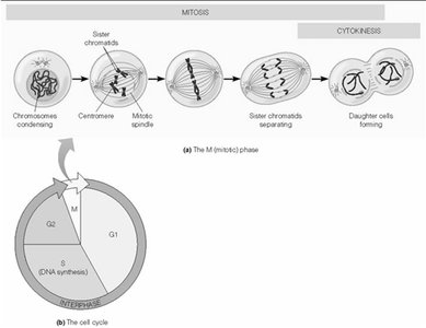
The eukaryotic cell cycle is a highly regulated series of events that leads to cell division and the production of two genetically identical daughter cells. It consists of interphase (G1, S, G2 phases) and the M phase (mitosis and cytokinesis).
G1 phase: Cell growth and preparation for DNA replication.
S phase: DNA synthesis, where chromosomes are replicated.
G2 phase: Further growth and preparation for mitosis.
M phase: Includes mitosis (nuclear division) and cytokinesis (cytoplasmic division).
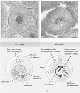
DNA Packaging: Chromatin and Chromosomes
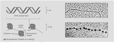
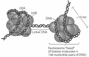
Nucleosomes and Chromatin Structure
DNA in eukaryotic cells is packaged into chromatin, which allows long DNA molecules to fit within the nucleus and plays a role in gene regulation. The basic unit of chromatin is the nucleosome.
Nucleosome: Consists of 146 base pairs of DNA wrapped around a core of eight histone proteins (two each of H2A, H2B, H3, and H4).
Linker DNA: The stretch of DNA between nucleosomes, associated with histone H1.
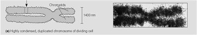
Chromatin condensation: During mitosis, chromatin condenses to form visible chromosomes, each consisting of two sister chromatids.
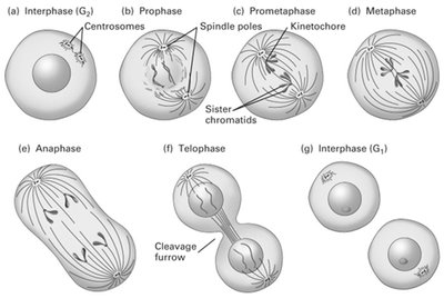
Mitosis: Phases and Key Events
Phases of Mitosis
Mitosis is the process by which a eukaryotic cell separates its duplicated chromosomes into two identical sets. It is divided into several distinct phases:
Prophase: Chromatin condenses into chromosomes; centrosomes move apart; mitotic spindle begins to form.
Prometaphase: Nuclear envelope breaks down; spindle microtubules attach to kinetochores on chromosomes.
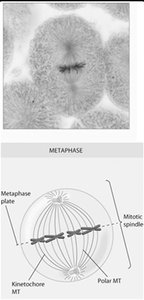
Metaphase: Chromosomes align at the metaphase plate; spindle fibers attach to kinetochores.
Anaphase: Sister chromatids separate and move toward opposite poles.
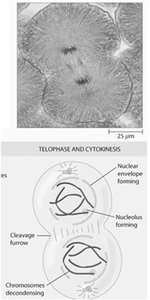
Telophase: Nuclear envelopes reform around the two sets of chromosomes; chromosomes decondense.
Cytokinesis: Division of the cytoplasm, resulting in two daughter cells.
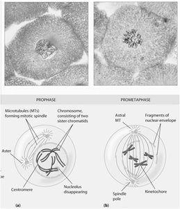
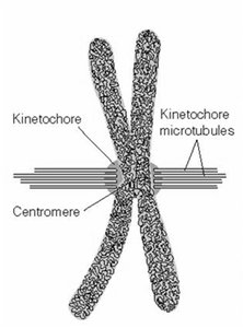
Centromere and Kinetochore
The centromere is the constricted region of a chromosome where sister chromatids are joined. The kinetochore is a protein complex assembled on the centromere, serving as the attachment site for spindle microtubules.
Centromere: Contains repetitive DNA sequences (CEN sequences).
Kinetochore: Plate-like structure that interacts with spindle microtubules to ensure proper chromosome segregation.
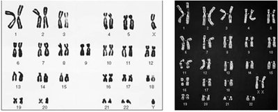
Metaphase and Karyotyping
During metaphase, chromosomes are maximally condensed and aligned at the metaphase plate. This stage is ideal for karyotyping, a technique used to visualize and analyze the number and structure of chromosomes.
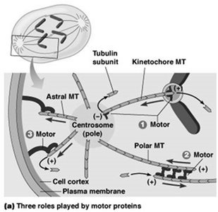
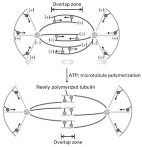
Kinetochore microtubules: Attach chromosomes to spindle poles.
Polar microtubules: Overlap at the cell center and help push the poles apart.
Astral microtubules: Radiate outward from the poles toward the cell cortex.
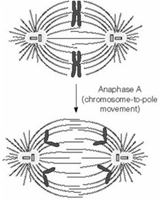
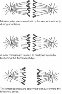
Anaphase and Chromosome Movement
Anaphase is characterized by the separation of sister chromatids, which are pulled toward opposite spindle poles. This movement is driven by the shortening of kinetochore microtubules and the action of motor proteins.
Anaphase A: Chromosomes move toward the poles as kinetochore microtubules shorten.
Anaphase B: Spindle poles move apart, further separating the chromosomes.
Motor proteins: Kinesins and dyneins facilitate microtubule dynamics and chromosome movement.


Telophase and Cytokinesis
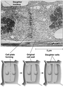
During telophase, the nuclear envelope reforms around the separated chromosomes, which decondense. Cytokinesis follows, dividing the cytoplasm and completing cell division.
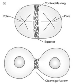
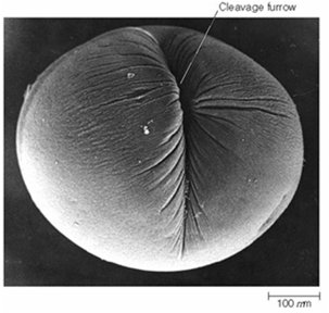
Contractile ring: Composed of actin and myosin II, forms beneath the plasma membrane at the metaphase plate and constricts to form the cleavage furrow.
Cytokinesis in animal cells: Involves the formation of a cleavage furrow.
Cytokinesis in plant cells: Involves the formation of a cell plate.
Cell Cycle Regulation
Checkpoints and Regulatory Molecules
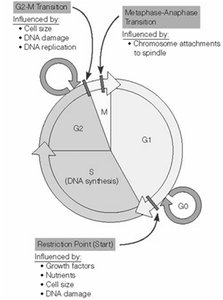
Progression through the cell cycle is tightly regulated by checkpoints that ensure each phase is completed correctly before the next begins. Key regulatory molecules include cyclin-dependent kinases (Cdks) and cyclins.
G1/S checkpoint (Restriction point): Checks for cell size, nutrients, growth factors, and DNA damage.
G2/M checkpoint: Ensures DNA replication is complete and checks for DNA damage.
Metaphase-Anaphase checkpoint: Ensures all chromosomes are properly attached to the spindle.
Cyclin-dependent kinases (Cdks): Protein kinases that require binding to cyclins for activation.
Cyclins: Regulatory proteins whose levels fluctuate during the cell cycle.

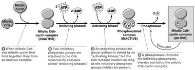
Activation and Inhibition of Cdks
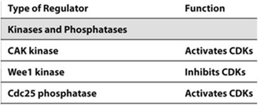
Cdk activity is regulated by cyclin binding, phosphorylation, and dephosphorylation. Specific kinases and phosphatases add or remove phosphate groups to control Cdk activity.
CAK kinase: Activates Cdks by phosphorylation.
Wee1 kinase: Inhibits Cdks by adding inhibitory phosphates.
Cdc25 phosphatase: Activates Cdks by removing inhibitory phosphates.

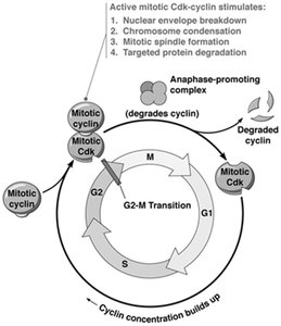
Maturation Promoting Factor (MPF) and the Anaphase-Promoting Complex (APC)
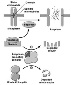
MPF is a cyclin-Cdk complex that triggers entry into M phase. The APC is a ubiquitin ligase that marks specific proteins for degradation, allowing progression from metaphase to anaphase and exit from mitosis.
MPF (Maturation-Promoting Factor): Composed of cyclin B and Cdk1; initiates mitosis by phosphorylating target proteins.
APC (Anaphase-Promoting Complex): Triggers the degradation of securin, activating separase to cleave cohesin and allow sister chromatid separation.
Mitotic cyclin degradation: Necessary for exit from mitosis and cytokinesis.
G1/S Transition and Tumor Suppressors
The G1/S transition is a critical control point regulated by the retinoblastoma protein (Rb) and the tumor suppressor p53. Loss of function in these proteins can lead to uncontrolled cell division and cancer.
Rb protein: Inhibits E2F transcription factor, preventing S phase entry in the absence of growth signals.
p53 protein: Activates DNA repair or apoptosis in response to DNA damage; loss of p53 function is common in cancers.
Checkpoint | Main Regulator | Function |
|---|---|---|
G1/S | Rb, Cyclin D/Cdk4/6 | Prevents S phase entry without growth signals |
G2/M | Cyclin B/Cdk1 (MPF) | Ensures DNA replication is complete |
Metaphase-Anaphase | APC | Ensures proper chromosome attachment and separation |
Example: Mutation in the Rb gene leads to retinoblastoma, a type of eye tumor, due to loss of cell cycle control.
Example: Mutation in p53 allows cells with DNA damage to proliferate, contributing to cancer development.
