 Back
BackThe Structural Basis of Cellular Information: DNA, Chromosomes, and the Nucleus
Study Guide - Smart Notes
 Tailored notes based on your materials, expanded with key definitions, examples, and context.
The Structural Basis of Cellular Information
Introduction to Genetic Material
Cells contain a set of instructions that determine their structure, function, and regulation. These instructions are encoded in genes, which are units of hereditary information passed from one generation to the next. The molecular basis of these instructions is found in nucleic acids, primarily DNA, and in some viruses, RNA.
Genes: Segments of DNA that encode functional products, usually proteins.
Hereditary Transmission: Genes are faithfully transmitted to daughter cells during cell division.
Flow of Genetic Information
Genetic information flows from DNA to RNA to protein, a process known as the central dogma of molecular biology. This flow occurs through two main processes: transcription and translation.
DNA Replication: Duplication of DNA before cell division.
Transcription: Synthesis of RNA from a DNA template.
Translation: Synthesis of proteins using mRNA as a template.

Chemical Nature of the Genetic Material
Discovery of DNA and Chromosomes
DNA was first isolated by Johann Friedrich Miescher in 1869. Chromosomes, the carriers of genetic material, were observed by Walther Flemming during cell division. Initially, proteins were thought to be the genetic material due to their complexity, but experiments later confirmed DNA's role.
RNA as Genetic Material in Some Viruses
While DNA is the genetic material in most organisms, some viruses use RNA. For example, the tobacco mosaic virus (TMV) contains RNA as its genetic material, and retroviruses convert their RNA genomes into DNA via reverse transcription.
DNA Structure and Properties
Chargaff’s Rules
Erwin Chargaff discovered that in DNA, the amount of adenine (A) equals thymine (T), and the amount of guanine (G) equals cytosine (C). These findings, known as Chargaff’s rules, were critical for understanding DNA structure.
The Double-Helix Model
DNA is a double-stranded, antiparallel polymer of deoxyribonucleotides. The two strands are complementary, with specific base pairing (A with T, G with C). The double helix allows for the faithful replication of genetic information.
Antiparallel Strands: The two DNA strands run in opposite directions (5' to 3' and 3' to 5').
Base Pairing: Purines (A, G) pair with pyrimidines (T, C).
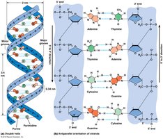
Key Features of DNA Structure
Major and Minor Grooves: The twisting of DNA creates grooves that are important for protein binding.
Phosphodiester Bonds: Link the 5' carbon of one nucleotide to the 3' carbon of the next.
DNA Length: Measured in base pairs (bp) or kilobases (kb).
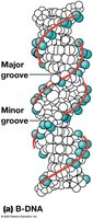
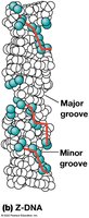
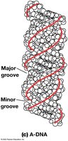
Helical Variants of Nucleic Acids
DNA can exist in several helical forms:
B-DNA: The most common, right-handed helix with clear major and minor grooves.
Z-DNA: Left-handed helix, less common, significance unclear.
A-DNA: Right-handed helix, favored by double-stranded RNA.
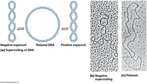
Supercoiling of DNA
DNA can be further twisted to form supercoils, which help compact the molecule. Positive supercoiling twists DNA in the same direction as the helix, while negative supercoiling twists it in the opposite direction.
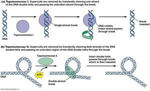
Topoisomerases
Topoisomerases are enzymes that regulate DNA supercoiling:
Type I Topoisomerases: Introduce transient single-strand breaks.
Type II Topoisomerases: Introduce double-strand breaks (e.g., DNA gyrase in bacteria).
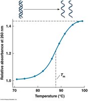
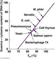
Denaturation and Renaturation of DNA
DNA strands can be separated (denatured) by heat or pH changes and can reanneal (renature) when conditions return to normal. The melting temperature (Tm) is the temperature at which half the DNA is denatured and depends on the GC content.
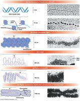
DNA Packaging in Cells
Chromatin and Nucleosomes
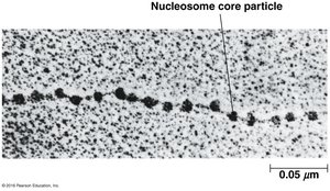
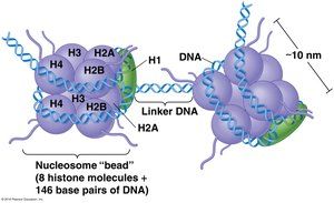
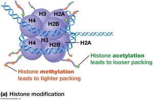
In eukaryotes, DNA is packaged with proteins to form chromatin. The basic unit of chromatin is the nucleosome, which consists of DNA wrapped around a histone octamer.
Histones: Small, basic proteins (H1, H2A, H2B, H3, H4) that facilitate DNA packaging.
Nucleosome Core Particle: 146 bp of DNA wrapped around a histone octamer.
Linker DNA: DNA between nucleosomes, associated with histone H1.
Higher-Order Chromatin Structure
Nucleosomes are further packed into 30-nm chromatin fibers, which are then organized into loops and scaffolds to form chromosomes.
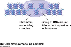
Chromatin Remodeling and Histone Modification
Chromatin structure can be dynamically regulated by chemical modifications of histones (e.g., methylation, acetylation) and by chromatin remodeling complexes (e.g., SWI/SNF family).
Methylation: Can activate or repress transcription depending on the site.
Acetylation: Generally associated with transcriptional activation and looser chromatin.
Euchromatin and Heterochromatin
Chromatin exists in two main forms:
Euchromatin: Less condensed, transcriptionally active.
Heterochromatin: Highly condensed, transcriptionally inactive. Includes constitutive (structural, e.g., centromeres, telomeres) and facultative (can switch to euchromatin) types.
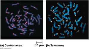
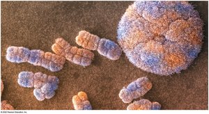
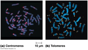
Centromeres and Telomeres
Centromeres are internal chromosome regions essential for proper segregation during cell division, while telomeres are repetitive sequences at chromosome ends that protect against degradation.

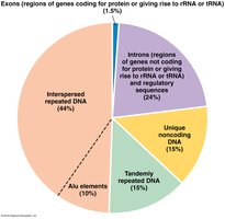
Repeated DNA Sequences
Tandemly and Interspersed Repeated DNA
Eukaryotic genomes contain large amounts of repeated DNA, which can be classified as tandemly repeated (adjacent repeats) or interspersed (scattered throughout the genome). Interspersed repeats include transposable elements such as LINEs and SINEs.
LINEs: Long interspersed nuclear elements, 6000–8000 bp, encode proteins for their own mobilization.
SINEs: Short interspersed nuclear elements, <500 bp, rely on other elements for movement (e.g., Alu sequences).
The Nucleus
Structure and Function
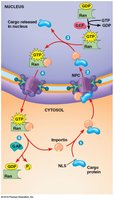
The nucleus is the site of DNA localization, replication, and transcription in eukaryotic cells. It is surrounded by a double-membrane nuclear envelope with nuclear pores that regulate molecular traffic.
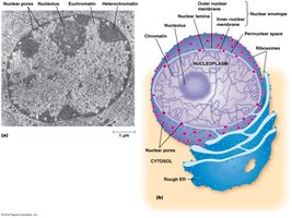
Nuclear Pore Complex (NPC)
The NPC is a large protein complex that facilitates selective transport of molecules between the nucleus and cytoplasm. Small molecules diffuse freely, while larger proteins and RNAs require active transport mediated by nuclear localization signals (NLS) and nuclear export signals (NES).
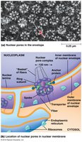
Nuclear Import and Export
Proteins with NLS are imported into the nucleus via the Ran/importin pathway, while RNAs are exported with the help of adaptor proteins and exportins. The Ran-GTP gradient across the nuclear envelope drives directionality of transport.

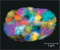
Nuclear Organization
The nucleus contains a nuclear matrix and lamina for structural support. Chromatin is organized into discrete chromosome territories. The nucleolus is the site of ribosome subunit assembly.
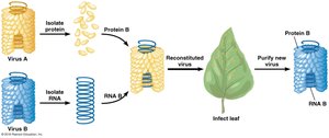
Experimental Evidence for DNA as Genetic Material
Griffith and Avery Experiments
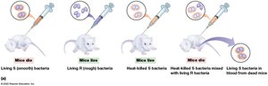
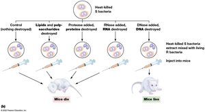
Frederick Griffith demonstrated genetic transformation in bacteria, showing that non-virulent strains could acquire virulence from dead pathogenic bacteria. Oswald Avery identified DNA as the transforming material responsible for this effect.
DNA Hybridization and Chromosome Identification
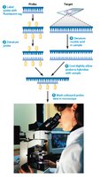
Fluorescent in Situ Hybridization (FISH)
FISH uses fluorescently labeled DNA probes to detect specific DNA sequences or chromosomes in cells, allowing for visualization of chromosomal abnormalities and gene mapping.
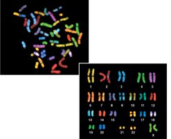
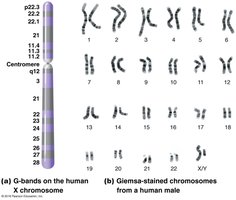
Chromosome Banding
Chromosomes can be distinguished by unique banding patterns produced by stains such as Giemsa, which reveal structural features and aid in karyotyping.
Summary Table: Major Types of Repeated DNA in Mammalian Genomes
Type | Description | Proportion of Genome |
|---|---|---|
Tandemly Repeated DNA | Multiple adjacent copies, usually short repeats | 10–15% |
Simple-Sequence Repeats | Tandem repeats <10 bp per repeat (satellite DNA) | Up to several hundred thousand copies |
Interspersed Repeated DNA | Dispersed, similar but not identical copies | 25–50% |
LINEs | Long interspersed nuclear elements (6000–8000 bp) | ~20% |
SINEs | Short interspersed nuclear elements (<500 bp, e.g., Alu) | ~10% |
