 Back
BackTrafficking Through the Endomembrane System: Protein Sorting, Vesicular Transport, and Cellular Digestion
Study Guide - Smart Notes
 Tailored notes based on your materials, expanded with key definitions, examples, and context.
Trafficking Through the Endomembrane System
Overview of Protein Trafficking
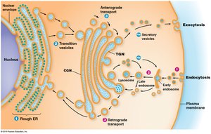
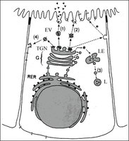
The endomembrane system is responsible for the synthesis, modification, sorting, and transport of proteins and lipids within eukaryotic cells. Proteins released into the endoplasmic reticulum (ER) lumen are routed to the Golgi complex, secretory vesicles, or returned to the ER. Sorting begins in the ER and early Golgi compartments, with mechanisms to retrieve or retain compartment-specific proteins. The final sorting of material destined to leave the Golgi occurs in the trans-Golgi network (TGN).
Protein Tags and Sorting Signals
Protein tags are molecular features that direct proteins to their correct cellular destinations. These tags may be amino acid sequences, hydrophobic domains, or oligosaccharide side chains. Tags can also exclude material from certain vesicles, ensuring specificity in protein sorting.
Retention Tags: Prevent proteins from leaving the ER (e.g., RXR sequence).
Retrieval Tags: Direct proteins back to the ER from the Golgi (e.g., KDEL or KKXX in mammals, HDEL in yeast).
When a protein with a retrieval tag binds its receptor, the complex is packaged into a transport vesicle for return to the ER.
Sorting of Golgi Complex Proteins
All Golgi-specific proteins are integral membrane proteins. The length of their hydrophobic membrane-spanning domains determines their localization within the Golgi cisternae. Membrane thickness increases from the ER to the plasma membrane, and proteins move until the membrane thickness exceeds their transmembrane domain length, blocking further migration.
Targeting of Soluble Lysosomal Proteins
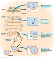
Soluble lysosomal enzymes are synthesized in the ER, undergo N-glycosylation, and are modified in the Golgi. Mannose residues on their oligosaccharide side chains are phosphorylated to form mannose-6-phosphate, which serves as a tag for delivery to lysosomes.
Insertion of Proteins into the ER Membrane
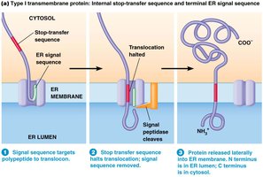
Stop-Transfer and Start-Transfer Sequences
Integral membrane proteins are synthesized and inserted into the ER membrane via two main mechanisms:
Stop-Transfer Sequences: Hydrophobic amino acid sequences that halt translocation through the ER membrane, anchoring the protein.
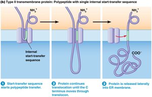
Internal Start-Transfer Sequences: Internal hydrophobic sequences that initiate membrane insertion without a typical N-terminal signal sequence.
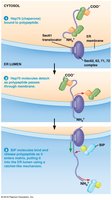
Posttranslational Import into the ER Lumen
Some proteins are synthesized in the cytosol and imported into the ER lumen posttranslationally. Chaperones such as Hsp70 keep the protein unfolded, and the Sec61 complex facilitates translocation. BiP, an ER chaperone, uses ATP hydrolysis to pull the polypeptide into the ER lumen.
Exocytosis and Endocytosis: Transport Across the Plasma Membrane
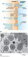
Exocytosis
Exocytosis is the process by which secretory vesicles fuse with the plasma membrane to release their contents outside the cell. This process is essential for secretion of proteins, neurotransmitters, and other molecules.
Constitutive Secretion: Continuous, unregulated release (e.g., mucus secretion).
Regulated Secretion: Vesicles fuse with the membrane in response to specific signals (e.g., neurotransmitter release).
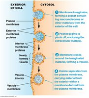
Endocytosis
Endocytosis is the process by which cells internalize extracellular materials by forming vesicles from the plasma membrane. There are several forms:
Phagocytosis: Uptake of solid particles.
Pinocytosis: Uptake of liquids.
Receptor-Mediated Endocytosis: Specific uptake of macromolecules via cell surface receptors.
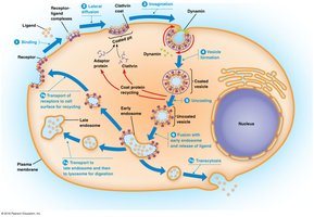
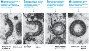
Receptor-Mediated Endocytosis
Cells use receptors to internalize specific ligands. Receptor-ligand complexes accumulate in coated pits, which invaginate and pinch off to form coated vesicles. Clathrin, adaptor proteins, and dynamin are essential for this process.
Coated Vesicles in Cellular Transport
Types of Coated Vesicles
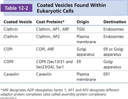
Coated vesicles are involved in protein and lipid transport throughout the endomembrane system. The main types are clathrin-coated, COPI-coated, and COPII-coated vesicles, each with distinct roles and protein compositions.
Coated Vesicle | Coat Proteins | Origin | Destination |
|---|---|---|---|
Clathrin | Clathrin, AP1, ARF | TGN | Endosomes |
Clathrin | Clathrin, AP2 | Plasma membrane | Endosomes |
COPI | COPI, ARF | Golgi apparatus | ER or Golgi apparatus |
COPII | COPII (Sec13/31 and Sec23/24), Sar1 | ER | Golgi apparatus |
Caveolin | Caveolin | Plasma membrane | ER? |
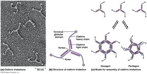
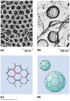
Clathrin-Coated Vesicles
Clathrin-coated vesicles are surrounded by a lattice of clathrin and adaptor proteins. The basic unit is the triskelion, which assembles into hexagons and pentagons to form a spherical vesicle.
COPI- and COPII-Coated Vesicles
COPI-coated vesicles mediate retrograde transport from the Golgi to the ER, while COPII-coated vesicles mediate anterograde transport from the ER to the Golgi. Both processes require small GTP-binding proteins (ARF for COPI, Sar1 for COPII) for coat assembly.
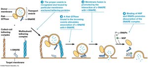
Vesicle Targeting and Fusion
SNARE Proteins and Vesicle Fusion
Vesicle targeting and fusion are mediated by SNARE proteins. v-SNAREs are found on vesicles, and t-SNAREs are found on target membranes. Rab GTPases promote SNARE association, while NSF and SNAPs are required for SNARE complex dissociation. Tethering proteins act over longer distances to facilitate vesicle recognition and docking.
Lysosomes and Cellular Digestion
Structure and Function of Lysosomes
Lysosomes are acidic organelles containing digestive enzymes (acid hydrolases) capable of degrading all major classes of biological macromolecules. Lysosomal enzymes are synthesized in the ER, delivered to endosomes, and activated as the environment acidifies.
pH: Maintained at 4.0–5.0 by ATP-dependent proton pumps (V-ATPases).
Development: Endosomes mature into lysosomes as they acquire enzymes and acidify.
Lysosomal Storage Diseases
Lysosomal storage diseases are characterized by the accumulation of indigestible material due to absent or defective lysosomal proteins. Examples include Type II glycogenosis, Hurler syndrome, Hunter syndrome, and Tay-Sachs disease.
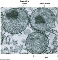
Peroxisomes
Structure and Function
Peroxisomes are single-membrane-bound organelles containing catalase for degrading hydrogen peroxide (H2O2). They are involved in hydrogen peroxide metabolism, detoxification, fatty acid oxidation, and catabolism of unusual substances.
Special Topics
Autophagy and Cancer
Autophagy is the process by which cells degrade and recycle their own components. It is essential for cellular homeostasis and has been linked to cancer development and progression.
Polarized Secretion
Polarized secretion refers to the targeted exocytosis of proteins to a specific surface of a cell, such as the secretion of digestive enzymes by intestinal cells only on the side facing the intestinal lumen.
Additional info:
Many receptor proteins, such as G protein-coupled receptors (GPCRs), have seven membrane-spanning domains, inserted by alternating start- and stop-transfer sequences.
Chaperones such as Hsp70 and BiP are essential for proper protein folding and translocation into the ER.
