 Indietro
Action Potentials and Neural Conduction: Study Notes
Guida di studio - Note intelligenti
 Appunti personalizzati basati sui tuoi materiali, ampliati con definizioni chiave, esempi e contesto.
Action Potentials
Introduction to Action Potentials
An action potential is a rapid, uniform depolarization and repolarization of the membrane potential of a cell, essential for communication in the nervous and muscular systems. In neurons, action potentials allow signals to travel along axons to other neurons, muscle fibers, or glands. In muscle fibers, they trigger contraction.
Trigger zone: The initial segment of the axon where the action potential is initiated.
All-or-none principle: An action potential either occurs fully or not at all, depending on whether the threshold is reached.
States of Voltage-Gated Channels
Sodium and Potassium Channel States
Voltage-gated channels are crucial for generating and propagating action potentials. They are abundant in the axolemma of neurons.
Sodium (Na+) channels: Three states—resting, activated, inactivated.
Potassium (K+) channels: Two states—resting (closed), activated (open).
Sodium Channel States
Resting state: Inactivation gate open, activation gate closed.
Activated state: Both gates open, allowing Na+ influx.
Inactivated state: Inactivation gate closed, activation gate open.
Potassium Channel States
Resting state: Channel closed.
Activated state: Channel open, allowing K+ efflux.
Phases and Events of an Action Potential
Three Phases of an Action Potential
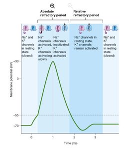
The action potential consists of three main phases, each defined by changes in membrane potential and channel activity.
Depolarization phase: Membrane potential rises toward zero and briefly becomes positive due to Na+ influx.
Repolarization phase: Membrane potential returns to a negative value as K+ exits the cell.
Hyperpolarization phase: Membrane potential becomes more negative than the resting potential before stabilizing.
Steps of an Action Potential
A local potential depolarizes the axolemma of the trigger zone to threshold.
Voltage-gated Na+ channels activate, Na+ enters, and the axon section depolarizes.
Na+ channels inactivate and voltage-gated K+ channels activate, initiating repolarization.
Na+ channels return to resting state, repolarization continues.
The axolemma may hyperpolarize before K+ channels return to resting state, then returns to resting membrane potential.
Refractory Periods
After an action potential, neurons experience a refractory period during which they are less responsive to stimuli.
Absolute refractory period: No additional stimulus can trigger another action potential, as Na+ channels are inactivated.
Relative refractory period: Only a strong stimulus can trigger another action potential; some K+ channels remain open, making depolarization more difficult.
Local Potentials vs. Action Potentials
Comparison of Local and Action Potentials
Local potentials: Graded, reversible, decremental (decrease over short distances).
Action potentials: Cause maximum, uniform depolarization (~+30 mV), irreversible, nondecremental (do not diminish in strength), travel long distances.
All-or-none principle: Action potentials only occur if threshold is reached; otherwise, no action potential is generated.
Propagation of Action Potentials
Mechanism of Propagation
Action potentials are self-propagating, meaning one action potential triggers another in an adjacent region of the axon, creating a domino effect. This process ensures signals travel in one direction—from the trigger zone to the axon terminals.
Depolarizing current from an action potential spreads through a section of the axolemma.
This current triggers adjacent voltage-gated Na+ channels to open, firing another action potential. The previously depolarized section repolarizes and becomes refractory.
The process repeats along the axon until the signal reaches the axon terminal.
Conduction Speed and Types
Factors Affecting Conduction Speed
Axon diameter: Larger axons conduct signals faster due to lower resistance.
Myelin sheath: Presence of myelin increases conduction speed.
Types of Conduction
Saltatory conduction: Occurs in myelinated axons. Action potentials "jump" from one node of Ranvier to the next, increasing speed and efficiency.
Continuous conduction: Occurs in unmyelinated axons. Each section of the axolemma must depolarize, resulting in slower signal transmission.
Example: Myelinated axons in the peripheral nervous system conduct signals much faster than unmyelinated axons due to saltatory conduction.
Clinical Application: Multiple Sclerosis (MS)
Multiple Sclerosis and Neural Conduction
Multiple Sclerosis (MS) is an autoimmune disorder in which immune cells attack the myelin sheath of axons in the central nervous system (CNS). This leads to progressive loss of myelin, reduced efficiency of saltatory conduction, and symptoms such as numbness, cognitive changes, and motor dysfunction.
Neuroglial cells affected: Oligodendrocytes (produce myelin in the CNS).
Conduction type affected: Saltatory conduction is impaired; conduction becomes more like continuous conduction.
Summary Table: Local vs. Action Potentials
Feature | Local Potentials | Action Potentials |
|---|---|---|
Amplitude | Graded (varies) | All-or-none (~+30 mV) |
Reversibility | Reversible | Irreversible |
Propagation | Decremental (fades) | Nondecremental (constant) |
Distance | Short | Long |
Key Terms and Definitions
Action potential: Rapid, uniform change in membrane potential.
Depolarization: Membrane potential becomes less negative.
Repolarization: Membrane potential returns to resting negative value.
Hyperpolarization: Membrane potential becomes more negative than resting.
Refractory period: Time during which a neuron is less responsive to stimuli.
Saltatory conduction: Fast conduction in myelinated axons.
Continuous conduction: Slow conduction in unmyelinated axons.
