 Indietro
Anatomy & Physiology II: The Cardiovascular and Respiratory Systems – Lab Study Guide
Guida di studio - Note intelligenti
 Appunti personalizzati basati sui tuoi materiali, ampliati con definizioni chiave, esempi e contesto.
The Cardiovascular System
Pathway of Blood Through the Heart and Body
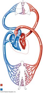
The cardiovascular system is responsible for the circulation of blood throughout the body, delivering oxygen and nutrients while removing waste products. The heart acts as a double pump, sending blood through two main circuits: the pulmonary circuit (to and from the lungs) and the systemic circuit (to and from the rest of the body).
Right Atrium: Receives deoxygenated blood from the body via the superior and inferior vena cava.
Tricuspid Valve: Prevents backflow as blood moves from the right atrium to the right ventricle.
Right Ventricle: Pumps deoxygenated blood through the pulmonary semilunar valve into the pulmonary trunk.
Pulmonary Trunk & Arteries: Carry blood to the lungs for oxygenation.
Lungs: Gas exchange occurs in the capillaries surrounding alveoli; blood becomes oxygenated.
Pulmonary Veins: Return oxygenated blood to the left atrium.
Left Atrium: Receives oxygenated blood from the lungs.
Bicuspid (Mitral) Valve: Prevents backflow as blood moves into the left ventricle.
Left Ventricle: Pumps oxygenated blood through the aortic semilunar valve into the aorta.
Aorta: Distributes oxygenated blood to the systemic circulation (all body tissues).
Systemic Capillaries: Exchange of gases, nutrients, and wastes with tissues.
Vena Cava: Returns deoxygenated blood to the right atrium, completing the circuit.
Key Concepts:
Blue indicates deoxygenated blood; red indicates oxygenated blood.
Valves ensure unidirectional flow and prevent backflow.
The pulmonary circuit is responsible for gas exchange in the lungs; the systemic circuit supplies the body.
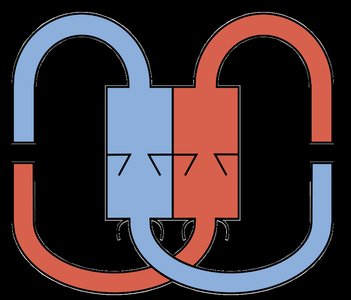
Additional info: The simplified diagram helps visualize the dual-circuit nature of the cardiovascular system, emphasizing the separation of oxygenated and deoxygenated blood.
The Respiratory System
Regions of the Pharynx
The pharynx is a muscular tube that serves as a passageway for both air and food. It is divided into three anatomical regions:
Nasopharynx: Located posterior to the nasal cavity; serves as an air passageway only.
Oropharynx: Posterior to the oral cavity; serves as a passageway for both air and food.
Laryngopharynx: Inferior region; leads to the larynx (air) and esophagus (food).
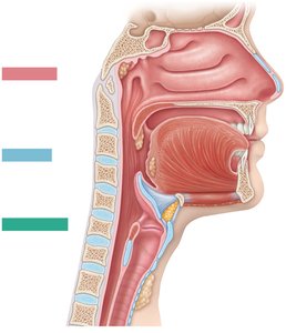
Key Terms: Nasopharynx, Oropharynx, Laryngopharynx
Anatomy of the Upper Respiratory Tract
The upper respiratory tract includes structures that filter, warm, and moisten incoming air. Key anatomical features include:
Nasal Cavity: Lined with mucosa and nasal conchae to increase surface area and turbulence.
Hard and Soft Palate: Separate the nasal cavity from the oral cavity.
Uvula: Prevents food from entering the nasopharynx during swallowing.
Tonsils: Lymphatic tissue (pharyngeal, palatine, lingual) that helps defend against pathogens.
Larynx: Contains the epiglottis (prevents food entry into the airway), vocal folds, and cartilages (thyroid, cricoid).
Trachea: Windpipe leading to the bronchi.
Esophagus: Posterior to the trachea; carries food to the stomach.
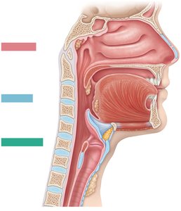
Additional info: The cribriform plate of the ethmoid bone allows passage of olfactory nerves; nasal conchae increase air turbulence and contact with mucosa.
Histology of the Trachea
The trachea is a flexible tube supported by C-shaped rings of hyaline cartilage. Its wall is composed of several layers:
Mucosa: Innermost layer, lined with pseudostratified ciliated columnar epithelium and goblet cells (produce mucus).
Submucosa: Connective tissue containing seromucous (tracheal) glands.
Hyaline Cartilage: Provides structural support and keeps the airway open.
Adventitia: Outermost connective tissue layer.
Trachealis Muscle: Smooth muscle connecting the open ends of the cartilage rings posteriorly, allowing flexibility and diameter adjustment.
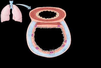
Key Terms: Pseudostratified ciliated columnar epithelium, seromucous glands, trachealis muscle, hyaline cartilage, mucosa, submucosa, adventitia
Structural and Functional Organization of the Respiratory System
The respiratory system can be organized both structurally and functionally:
Structural Organization:
Upper Respiratory Tract: Nose, nasal cavity, pharynx
Lower Respiratory Tract: Larynx, trachea, bronchi, bronchioles, lungs
Functional Organization:
Conducting Zone: All passageways that conduct air (nose to terminal bronchioles)
Respiratory Zone: Sites of gas exchange (respiratory bronchioles, alveolar ducts, alveoli)
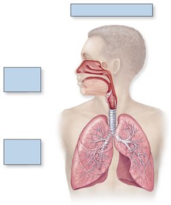
Additional info: The conducting zone filters, warms, and moistens air, while the respiratory zone is where oxygen and carbon dioxide are exchanged with blood.
The Bronchial Tree
The bronchial tree consists of branching airways that conduct air from the trachea into the lungs:
Main (Primary) Bronchi: Right and left branches from the trachea entering each lung.
Lobar (Secondary) Bronchi: Branches supplying each lobe of the lungs (3 right, 2 left).
Segmental (Tertiary) Bronchi: Further divisions supplying bronchopulmonary segments.
Smaller Bronchi and Bronchioles: Continue branching, ending in terminal bronchioles.
Key Terms: Larynx, trachea, main bronchus, lobar bronchus, segmental bronchus, left vs right lung
Anatomical Relationships in the Thoracic Cavity
The thoracic cavity contains the lungs, heart, and associated structures. Key anatomical relationships include:
Lungs: Divided into lobes (right: 3, left: 2) and surrounded by pleural membranes.
Pleura: Double-layered serous membrane (parietal pleura lines the cavity, visceral pleura covers the lungs).
Pleural Cavity: Space between pleural layers containing lubricating fluid.
Mediastinum: Central compartment containing the heart, trachea, esophagus, and major vessels.
Diaphragm: Muscle separating thoracic and abdominal cavities; primary muscle of respiration.
Additional info: The cardiac notch is a concavity in the left lung accommodating the heart.
Bronchioles and Alveoli
Gas exchange occurs in the respiratory zone, which includes:
Bronchioles: Small airways lacking cartilage; terminal bronchioles mark the end of the conducting zone.
Respiratory Bronchioles: First part of the respiratory zone; have some alveoli in their walls.
Alveolar Ducts and Alveoli: Alveoli are tiny air sacs where gas exchange occurs across simple squamous epithelium.
Pulmonary Capillaries: Surround alveoli, facilitating exchange of O2 and CO2 between air and blood.
Flow of Blood in Pulmonary Circuit: Right ventricle → pulmonary artery → pulmonary arteriole → pulmonary capillaries (gas exchange) → pulmonary venule → pulmonary vein → left atrium
Additional info: Alveolar pores connect adjacent alveoli, equalizing air pressure and providing alternate routes for air flow.
