 Indietro
Anatomy and Physiology of the Heart: Structure, Function, and Clinical Aspects
Guida di studio - Note intelligenti
 Appunti personalizzati basati sui tuoi materiali, ampliati con definizioni chiave, esempi e contesto.
Heart Anatomy and Circulation
Pulmonary and Systemic Circuits
The heart functions as a dual pump, circulating blood through two distinct circuits: the pulmonary and systemic circuits. The right side of the heart receives oxygen-poor blood from the body and pumps it to the lungs for gas exchange, while the left side receives oxygen-rich blood from the lungs and pumps it to the rest of the body.
Pulmonary Circuit: Right side pumps blood to lungs to eliminate CO2 and absorb O2.
Systemic Circuit: Left side pumps oxygenated blood to body tissues.
Receiving Chambers: Right atrium (systemic circuit), Left atrium (pulmonary circuit).
Pumping Chambers: Right ventricle (pulmonary circuit), Left ventricle (systemic circuit).
Size, Location, and Orientation of the Heart
The heart is roughly the size of a fist and weighs less than one pound. It is located in the mediastinum, between the second rib and fifth intercostal space, with two-thirds of its mass to the left of the midsternal line. The base leans toward the right shoulder, and the apex points toward the left hip.
Heart Structure and Coverings
Pericardium and Heart Wall Layers
The heart is enclosed by the pericardium, a double-walled sac consisting of a superficial fibrous layer and a deep serous layer. The serous pericardium has a parietal layer lining the fibrous pericardium and a visceral layer (epicardium) covering the heart. The pericardial cavity between these layers contains fluid to reduce friction.
Epicardium: Visceral layer of serous pericardium.
Myocardium: Bundles of contractile cardiac muscle cells; contains the cardiac skeleton for structural support.
Endocardium: Innermost layer, continuous with blood vessel endothelium.
Clinical Conditions
Pericarditis: Inflammation of the pericardium causing friction rub.
Cardiac Tamponade: Compression of the heart due to excess fluid in the pericardial cavity.
Chambers and Associated Great Vessels
Internal Features
The heart has four chambers: two atria and two ventricles, separated by septa. The right atrium receives blood from the body via the superior and inferior vena cava and the coronary sinus. The left atrium receives blood from the lungs via four pulmonary veins.
Interatrial Septum: Separates atria; contains fossa ovalis (remnant of fetal foramen ovale).
Interventricular Septum: Separates ventricles.
Auricles: Increase atrial volume.
Ventricles: Discharging chambers; right ventricle pumps to pulmonary trunk, left ventricle to aorta.
Trabeculae Carneae: Muscle ridges in ventricles.
Papillary Muscles: Anchor chordae tendineae attached to valves.
Heart Valves
Atrioventricular (AV) Valves
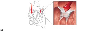
AV valves prevent backflow into the atria during ventricular contraction. The tricuspid valve is on the right, and the mitral (bicuspid) valve is on the left. Chordae tendineae anchor the valve cusps to papillary muscles, ensuring proper closure.
Tricuspid Valve: Right AV valve, three cusps.
Mitral Valve: Left AV valve, two cusps.
Chordae Tendineae: Prevent valve eversion.
Semilunar (SL) Valves
SL valves prevent backflow from arteries into ventricles. The pulmonary SL valve is between the right ventricle and pulmonary trunk; the aortic SL valve is between the left ventricle and aorta.
Three Cusps: Each SL valve has three half-moon-shaped cusps.
Clinical Valve Disorders
Incompetent Valve: Allows backflow; heart repumps same blood.
Valvular Stenosis: Stiff flaps restrict opening; heart must exert more force.
Valve Replacement: Mechanical, animal, or cadaver valves.
Pathway of Blood Through the Heart
Right Side
Blood enters via SVC, IVC, and coronary sinus → right atrium → tricuspid valve → right ventricle → pulmonary semilunar valve → pulmonary trunk → pulmonary arteries → lungs.
Left Side
Blood enters via four pulmonary veins → left atrium → mitral valve → left ventricle → aortic semilunar valve → aorta → systemic circulation.
Ventricular Anatomy and Function
Left ventricle walls are three times thicker than right; pumps with greater pressure.
Pulmonary circuit is short, low-pressure; systemic circuit is long, high-friction.
Coronary Circulation
Blood Supply to the Heart
Coronary circulation delivers 1/20th of the body's blood supply to the heart muscle, primarily during relaxation. The left ventricle receives most of the supply. Coronary arteries branch from the aorta and form anastomoses for collateral circulation.
Angina Pectoris: Temporary deficiency in blood delivery; causes thoracic pain.
Myocardial Infarction: Prolonged blockage; cell death replaced by scar tissue.
Microscopic Anatomy of Cardiac Muscle
Cardiac Muscle Fibers
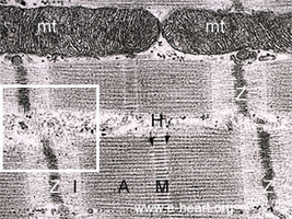
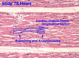
Cardiac muscle cells are striated, short, branched, and interconnected, typically with one central nucleus. They contain numerous large mitochondria for resistance to fatigue and are organized into sarcomeres with Z discs, A bands, and I bands.
Intercalated Discs: Junctions containing desmosomes (mechanical strength) and gap junctions (electrical coupling).
Endomysium: Connective tissue matrix with capillaries, linking muscle to cardiac skeleton.
Comparison: Skeletal vs. Cardiac Muscle
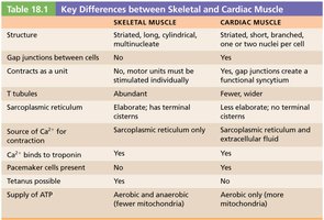
Key differences between skeletal and cardiac muscle are summarized below:
Feature | Skeletal Muscle | Cardiac Muscle |
|---|---|---|
Structure | Striated, long, cylindrical, multinucleate | Striated, short, branched, one or two nuclei per cell |
Gap Junctions | No | Yes |
Contracts as Unit | No | Yes |
T tubules | Abundant | Less elaborate |
Source of Ca2+ | Sarcoplasmic reticulum only | Sarcoplasmic reticulum and extracellular fluid |
Pacemaker Cells | No | Yes |
Tetanus Possible | Yes | No |
ATP Supply | Aerobic and anaerobic | Aerobic only (more mitochondria) |
Electrical Events of the Heart
Intrinsic Cardiac Conduction System
The heart depolarizes and contracts without nervous system stimulation, thanks to the intrinsic conduction system composed of autorhythmic cells. The sequence of excitation ensures coordinated contraction.
Sinoatrial (SA) Node: Pacemaker; initiates impulses.
Atrioventricular (AV) Node: Delays impulse, allowing atrial contraction.
AV Bundle (Bundle of His): Only electrical connection between atria and ventricles.
Bundle Branches: Carry impulses toward apex.
Purkinje Fibers: Depolarize ventricular muscle.
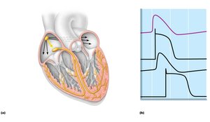
Action Potential in Cardiac Muscle
Cardiac muscle action potentials feature a plateau phase, resulting in longer contraction and refractory periods compared to skeletal muscle. This prevents tetanic contractions and ensures efficient blood ejection.
Electrocardiography (ECG/EKG)
ECG Features
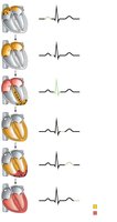
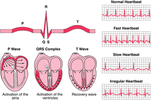
An ECG records the electrical activity of the heart. Key features include:
P wave: Depolarization of SA node and atria.
QRS complex: Ventricular depolarization and atrial repolarization.
T wave: Ventricular repolarization.
P-R interval: Atrial to ventricular excitation.
S-T segment: Ventricular myocardium depolarized.
Q-T interval: Ventricular depolarization through repolarization.
Mechanical Events of the Heart
Cardiac Cycle
The cardiac cycle consists of systole (contraction) and diastole (relaxation), representing a complete heartbeat. Mechanical events follow electrical events seen on ECG.
Ventricular Filling: Blood flows passively into ventricles; atrial contraction completes filling.
Ventricular Systole: Ventricles contract; AV valves close, SL valves open for ejection.
Isovolumetric Relaxation: Ventricles relax; SL valves close, AV valves open when atrial pressure exceeds ventricular.
Cardiac Output and Regulation
Cardiac Output (CO)
Cardiac output is the volume of blood pumped by each ventricle per minute.
Formula:
Stroke Volume (SV):
Normal Values: At rest, CO ≈ 5.25 L/min; can increase to 20–25 L/min in nonathletes, up to 35 L/min in athletes.
Regulation of Stroke Volume
Preload: Degree of stretch before contraction; Frank-Starling law.
Contractility: Strength at given muscle length; increased by sympathetic stimulation and certain hormones.
Afterload: Pressure ventricles must overcome; increased in hypertension.
Regulation of Heart Rate
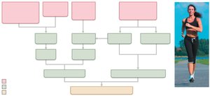
Autonomic Nervous System: Sympathetic increases HR and contractility; parasympathetic decreases HR.
Chemical Regulation: Hormones (epinephrine, thyroxine) and ions (Ca2+, K+).
Other Factors: Age, gender, exercise, body temperature.
Clinical Aspects and Homeostatic Imbalances
ECG Abnormalities and Arrhythmias
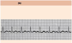
Arrhythmias: Irregular heart rhythms; may cause fibrillation.
Heart Block: Defective AV node; treated with artificial pacemaker.
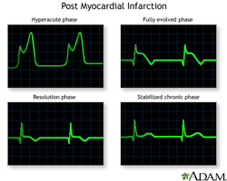
ECG Changes: Enlarged R waves, S-T segment changes, prolonged Q-T interval indicate pathology.
Heart Sounds and Murmurs
Lub-dup: AV and SL valve closure.
Heart Murmurs: Abnormal sounds due to valve problems.
Congestive Heart Failure (CHF)
Causes: Coronary atherosclerosis, high blood pressure, multiple infarcts, dilated cardiomyopathy.
Left-sided Failure: Pulmonary congestion.
Right-sided Failure: Peripheral congestion.
Treatment: Fluid removal, drugs to reduce afterload and increase contractility.
Congenital Heart Defects
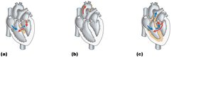
Types: Mixing of oxygen-poor and oxygen-rich blood (septal defects), narrowed valves/vessels (coarctation of aorta), Tetralogy of Fallot (multiple defects).
Treatment: Surgical correction.
Age-Related Changes Affecting the Heart
Sclerosis and Thickening: Valve flaps, leading to murmurs.
Decline in Cardiac Reserve: Reduced efficiency.
Fibrosis: Stiffened heart, arrhythmias.
Atherosclerosis: Preventable with healthy lifestyle.
Additional info: Regular exercise and a healthy diet are important for maintaining heart function and preventing age-related decline.
