 Indietro
Anatomy and Physiology of the Heart: Structure, Function, and Conducting System
Guida di studio - Note intelligenti
 Appunti personalizzati basati sui tuoi materiali, ampliati con definizioni chiave, esempi e contesto.
Anatomy of the Heart
Location and Morphological Features
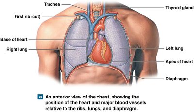
The heart is a muscular organ located in the mediastinum, the central compartment of the thoracic cavity, between the two pleural cavities. It is positioned posterior to the sternum and anterior to the vertebral column, resting on the diaphragm. - Base of the heart: Superior portion, where major vessels enter and exit. - Apex of the heart: Inferior, pointed end directed downward and to the left. - Surrounding structures: Lungs, ribs, and diaphragm. 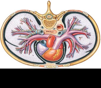
Pericardium and Layers of the Heart
The heart is enclosed by the pericardium, a double-walled sac that protects and anchors the heart. - Fibrous pericardium: Tough, outer layer providing protection and anchorage. - Serous pericardium: Inner layer, divided into: - Parietal layer: Lines the internal surface of the fibrous pericardium. - Visceral layer (epicardium): Covers the heart surface. - Pericardial cavity: Space between parietal and visceral layers, filled with pericardial fluid to reduce friction. 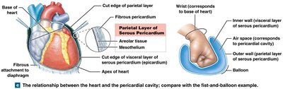
Layers of the Heart Wall
The heart wall consists of three distinct layers: - Epicardium: Outer layer, also known as visceral pericardium. - Myocardium: Middle layer, composed of cardiac muscle responsible for contraction. - Endocardium: Inner layer, lining the heart chambers and valves. 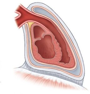
Cardiac Chambers and Blood Flow
Four Chambers of the Heart
The heart contains four chambers: - Right atrium: Receives deoxygenated blood from the body. - Right ventricle: Pumps deoxygenated blood to the lungs. - Left atrium: Receives oxygenated blood from the lungs. - Left ventricle: Pumps oxygenated blood to the body. 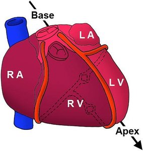
Blood Flow Through the Heart
Blood flows through the heart in a specific sequence: 1. Deoxygenated blood enters the right atrium from systemic veins. 2. Blood moves to the right ventricle, which pumps it to the lungs for oxygenation. 3. Oxygenated blood returns to the left atrium. 4. Blood is pumped from the left ventricle to the systemic circulation. - Atria: Thin-walled chambers for receiving blood. - Ventricles: Thick-walled chambers for pumping blood out. 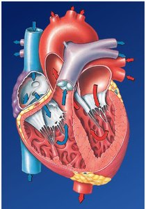
Conducting System of the Heart
Overview of the Conducting System
The heart's conducting system consists of specialized cardiac muscle cells that initiate and distribute electrical impulses, enabling coordinated contraction. - Autorhythmicity: Cardiac muscle contracts without external stimulation. - Pacemaker cells: Located in the sinoatrial (SA) node and atrioventricular (AV) node. - Conducting cells: Include internodal pathways, AV bundle, bundle branches, and Purkinje fibers. 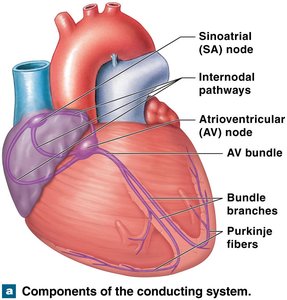
Impulse Conduction Pathway
The sequence of electrical conduction through the heart is as follows: 1. SA node activity: Initiates impulse and atrial activation. 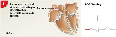 2. Impulse spreads: Across atria to AV node.
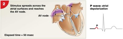 3. AV node delay: Allows atrial contraction.
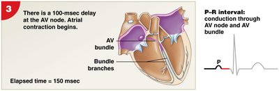 4. Impulse travels: Through AV bundle, bundle branches, and moderator band to Purkinje fibers.
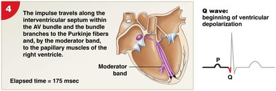 5. Purkinje fibers: Distribute impulse to ventricular myocardium, initiating ventricular contraction.
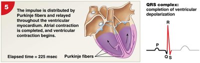
Electrocardiography (ECG/EKG)
Features of an ECG
An ECG records the electrical activity of the heart, displaying characteristic waves: - P wave: Atrial depolarization. - QRS complex: Ventricular depolarization; ventricles contract shortly after R wave. - T wave: Ventricular repolarization. 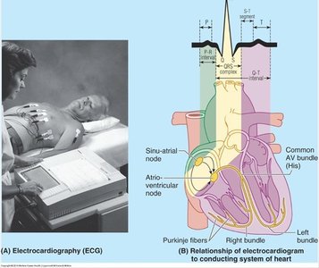
ECG Time Intervals
- P–R interval: Time from start of atrial depolarization to start of QRS complex. - Q–T interval: Time required for ventricles to undergo a single cycle of depolarization and repolarization. 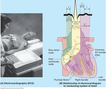
Coronary Circulation
Arterial Supply to the Heart
The heart receives blood through two main coronary arteries: - Right coronary artery - Left coronary artery These arteries supply the atria, ventricles, and conducting system (including SA and AV nodes). 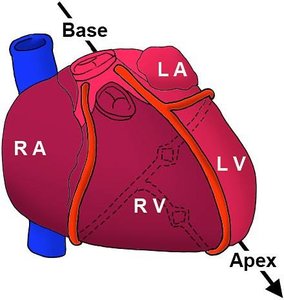
Coronary Heart Disease
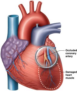
Partial or complete blockage of coronary arteries can prevent oxygen from reaching cardiac muscle, leading to reduced circulatory supply and potential myocardial infarction (heart attack). - Atherosclerotic plaque: Narrows arteries, restricting blood flow. - Occluded coronary artery: Causes damage to heart muscle. 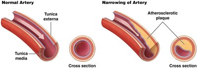
Summary Table: Heart Structure and Function
Structure | Function |
|---|---|
Right Atrium | Receives deoxygenated blood from body |
Right Ventricle | Pumps blood to lungs |
Left Atrium | Receives oxygenated blood from lungs |
Left Ventricle | Pumps blood to body |
Valves | Control blood flow direction |
SA Node | Initiates electrical impulse |
AV Node | Delays impulse for atrial contraction |
Coronary Arteries | Supply oxygen to heart muscle |
Key Points to Remember
Atria receive blood from veins.
Ventricles pump blood out of the heart.
Valves control blood flow via opening and closing.
Conducting system allows the heart to pump blood efficiently.
Conduction moves from the SA node through to the AV node.
Coronary artery disease prevents oxygen from reaching cardiac muscle.
