 Indietro
Anatomy and Physiology of the Reproductive System
Guida di studio - Note intelligenti
 Appunti personalizzati basati sui tuoi materiali, ampliati con definizioni chiave, esempi e contesto.
The Reproductive System
Overview and Functions
The reproductive system is essential for the continuation of the human species, comprising organs and structures responsible for producing gametes, secreting hormones, and facilitating fertilization and development. - Gonads: Organs that produce gametes (sperm or oocytes) and sex hormones. - Ducts: Transport gametes from gonads to the exterior or to sites of fertilization. - Accessory glands: Secrete fluids that support gametes and reproductive processes. - External genitalia: Structures visible externally, involved in copulation and protection.
Male Reproductive System
Anatomy and Major Structures
The male reproductive system is specialized for the production and delivery of sperm. Its main components include the testes, ducts, accessory glands, and external genitalia. - Testes: Male gonads that produce sperm and secrete androgens (male sex hormones). - Ducts: Include the epididymis, ductus deferens, ejaculatory duct, and urethra. - Accessory glands: Seminal glands, prostate, and bulbo-urethral glands produce seminal fluid. - External genitalia: Penis and scrotum. 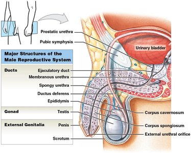
Epididymis
The epididymis is a coiled tube attached to the posterior border of each testis, serving as the site for sperm maturation and storage. - Head: Receives sperm from efferent ductules. - Body: Located on the posterior surface of the testis. - Tail: Primary storage site for sperm, connects to the ductus deferens. 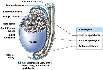
Ductus Deferens (Vas Deferens)
The ductus deferens is a muscular tube that transports sperm from the epididymis to the ejaculatory duct. - Length: 40–45 cm. - Pathway: Ascends through the inguinal canal, curves along the bladder, and joins the seminal gland. - Function: Stores sperm for several months in a low metabolic state. 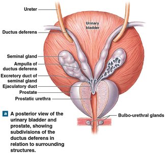
Accessory Glands
Accessory glands produce the fluid component of semen, each with unique biochemical properties. - Seminal glands: Contribute to the majority of seminal fluid. - Prostate: Secretes enzymes and nutrients. - Bulbo-urethral glands: Produce mucus for lubrication. 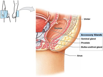
Penis and Erectile Tissues
The penis is the organ of copulation, containing erectile tissues that facilitate erection. - Corpora cavernosa: Paired columns of erectile tissue. - Corpus spongiosum: Surrounds the urethra and forms the glans penis. - Erection: Mediated by the parasympathetic nervous system, allowing blood to fill erectile tissues. 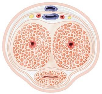
Female Reproductive System
Anatomy and Major Structures
The female reproductive system is designed for the production of oocytes, fertilization, and support of embryonic development. - Ovaries: Female gonads that produce oocytes and secrete hormones. - Uterine tubes: Transport oocytes from ovaries to uterus; site of fertilization. - Uterus: Muscular organ that supports embryo and fetus. - Vagina: Muscular tube connecting uterus to exterior; serves as birth canal. - External genitalia: Includes labia majora, labia minora, and vestibule. 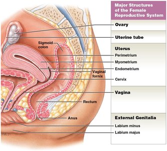
Gross Anatomy of the Uterus
The uterus is a thick-walled, muscular organ that facilitates the development of the embryo and fetus. - Cervix: Inferior opening into the vagina. - Uterine tubes: Project laterally and open near the ovaries. - Vagina: Passage for menses, birth canal, and receives penis during intercourse. 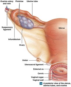
Layers of the Uterus
The uterine wall consists of three layers: - Perimetrium: Incomplete serous membrane, continuous with peritoneal lining. - Myometrium: Thick, muscular middle layer. - Endometrium: Thin, glandular inner layer, actively involved in the menstrual cycle. 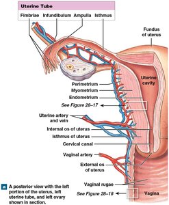
Uterine Tubes
Uterine tubes are divided into three segments: - Infundibulum: Funnel-shaped, with fimbriae extending into pelvic cavity. - Ampulla: Middle segment, muscle thickness increases near uterus. - Isthmus: Short segment adjacent to uterine wall. 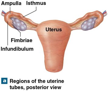
Ovaries
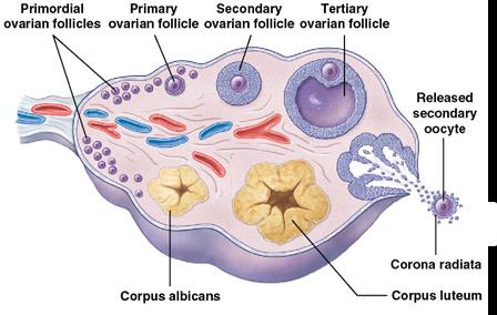
Ovaries are small, almond-shaped organs with three main functions: - Production of oocytes: Immature female gametes. - Secretion of hormones: Estrogens and progesterone. - Secretion of inhibin: Feedback control of pituitary FSH. 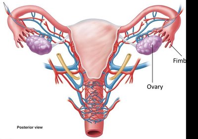
The Ovarian Cycle
Follicular Development
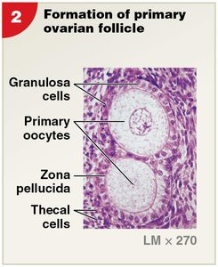
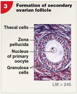
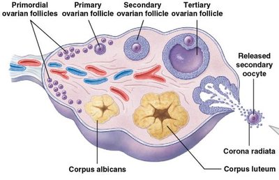
The ovarian cycle is a monthly process of follicle maturation, ovulation, and degeneration. - Primordial follicles: Contain primary oocytes in clusters called egg nests. - Primary follicle: Formed from activated primordial follicle. - Secondary follicle: Develops from primary follicle. - Tertiary follicle: Fluid accumulates, follicle enlarges. 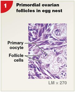
Phases of the Ovarian Cycle
The ovarian cycle is divided into two main phases: - Follicular phase: Growth and maturation of follicles; ends with ovulation. - Luteal phase: Formation and degeneration of corpus luteum; prepares uterus for pregnancy.
Ovulation and Corpus Luteum
- Ovulation: Release of secondary oocyte from tertiary follicle into pelvic cavity. - Corpus luteum: Formed from granulosa cells; secretes progesterone and estrogens. - Corpus albicans: Scar tissue formed from degenerated corpus luteum; marks end of cycle.
Uterine Cycle (Menstrual Cycle)
Phases of the Uterine Cycle
The uterine cycle is a repeating series of changes in the endometrium, lasting 21–35 days. - Menstrual phase: Shedding of functional endometrial layer. - Proliferative phase: Rapid growth of new endometrial tissue, stimulated by estrogen. - Secretory phase: Uterine glands secrete nutrients; occurs after ovulation.
Female External Genitalia
Vulva and Associated Structures
The vulva, or pudendum, includes the external genitalia and associated erectile tissues. - Labium minus: Thin folds of skin on either side of the midline. - Vestibule: Area between labium minus, containing openings of urethra and vagina. - Labium majus: Larger folds lateral to the vestibule.
Integration with Other Body Systems
Reproductive System Interactions
The reproductive system interacts with other body systems, including the endocrine, nervous, and circulatory systems, to regulate reproductive function and support pregnancy.
Summary Table: Major Structures of the Reproductive System
Male Structures | Female Structures |
|---|---|
Testis | Ovary |
Epididymis | Uterine tube |
Ductus deferens | Uterus |
Seminal gland | Vagina |
Prostate | Labium minus/majus |
Bulbo-urethral gland | Vestibule |
Penis | Clitoris |
Scrotum | Vulva |
Additional info: Academic context was added to clarify the functions and relationships of reproductive structures, and to provide a self-contained study guide suitable for exam preparation.
