 Indietro
Anatomy & Physiology Study Guide: Chapter 9 – Joints
Guida di studio - Note intelligenti
 Appunti personalizzati basati sui tuoi materiali, ampliati con definizioni chiave, esempi e contesto.
Joints (Articulations)
Definition and Importance
Joints, also known as articulations, are the locations where two bones connect. Because bones are rigid and relatively inflexible, movement in the body occurs primarily at joints. Some joints allow movement, while others are immobile, serving to strongly connect bones.
Joint: The site where two bones meet.
Articulation: Another term for joint.
Movement: Occurs only at joints due to the rigidity of bones.
Structural and Functional Classification of Joints
Joints are classified both structurally and functionally. Structural classification is based on the material connecting the bones, while functional classification is based on the degree of movement permitted.
Structural Classifications: Fibrous, Cartilaginous, Bony fusion, Synovial.
Functional Classifications: Synarthrosis (immobile), Amphiarthrosis (slightly movable), Diarthrosis (freely movable).
Structural Classification of Joints
Fibrous Joints
Fibrous joints are connected by fibrous connective tissue and generally allow little to no movement.
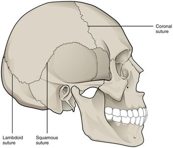
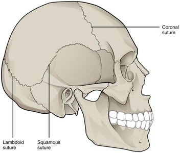
Suture: Found between bones of the skull.
Gomphosis: Binds teeth to bony sockets via the periodontal ligament.
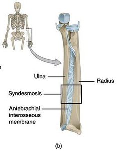
Syndesmosis: Bones connected by a ligament or interosseous membrane (e.g., between radius and ulna).
Cartilaginous Joints
Cartilaginous joints are connected by cartilage and allow limited movement.
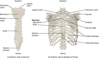
Synchondrosis: A rigid, cartilaginous bridge (e.g., first pair of vertebrosternal ribs and sternum).
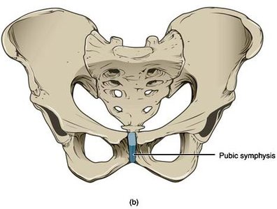
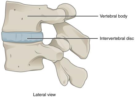
Symphysis: Bones separated by a pad of fibrocartilage (e.g., pubic symphysis, intervertebral discs).
Bony Fusion (Synostosis)
Synostosis is a totally rigid, immovable joint formed when two bones fuse and the boundary disappears (e.g., epiphyseal lines of mature long bones). 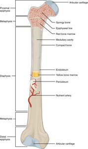
Synovial Joints
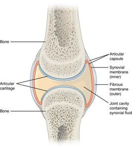
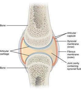
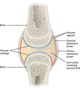
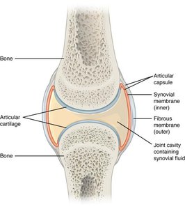
Synovial joints are characterized by a fluid-filled joint cavity and permit a wide range of motion.
Articular capsule: Surrounds the joint, composed of fibrous connective tissue.
Synovial membrane: Secretes synovial fluid for lubrication, nutrient supply, and shock absorption.
Articular cartilage: Covers the articulating surfaces, reduces friction.
Functional Classification of Joints
Synarthrosis (Immobile Joints)
Synarthroses are immobile joints that strongly connect two bones. Examples include skull sutures, gomphoses, synchondroses, and synostoses.
Fibrous synarthrosis: Sutures, gomphoses.
Cartilaginous synarthrosis: Synchondrosis.
Bony synarthrosis: Synostosis.
Amphiarthrosis (Slightly Movable Joints)
Amphiarthroses permit limited movement and are stronger than diarthroses. Examples include syndesmoses and symphyses.
Fibrous amphiarthrosis: Syndesmosis (e.g., interosseous membranes).
Cartilaginous amphiarthrosis: Symphysis (e.g., pubic symphysis, intervertebral discs).
Diarthrosis (Freely Movable Joints)
Diarthroses, or synovial joints, permit a wide range of motion. Examples include shoulder, elbow, hip, wrist, and ankle joints.
Synovial joints: Characterized by a fluid-filled cavity.
Classification of Synovial Joints by Axis of Motion
Uniaxial Joints
Allow movement in one plane (single axis). Examples: elbow and ankle joints. 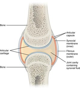
Biaxial Joints
Allow movement in two planes (two axes). Examples: wrist and knuckle joints. 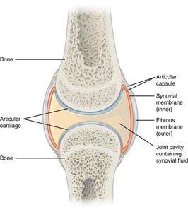
Triaxial Joints
Allow movement in three planes (three axes). Examples: hip and shoulder joints. 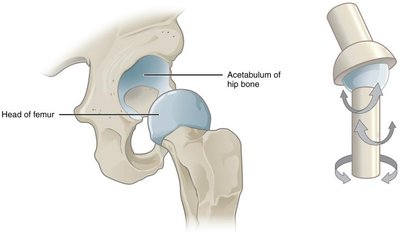
Synovial Joint Structure and Accessory Components
Articular Capsule and Synovial Membrane
The articular capsule surrounds the synovial joint and attaches to the periosteum of each bone. Deep to the capsule is the synovial membrane, which secretes synovial fluid.
Synovial fluid: Lubricates, nourishes, and absorbs shock.
Articular Cartilage
Covers the articulating surfaces, is similar to hyaline cartilage but contains more water and lacks a perichondrium.
Accessory Ligaments
Ligaments support, strengthen, and reinforce synovial joints.
Extracapsular ligaments: Outside the capsule.
Intracapsular ligaments: Inside the capsule.
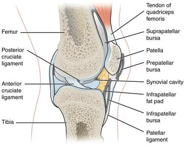
Meniscus
A pad of fibrocartilage found in some synovial joints, such as the knee, providing shock absorption and cushioning. 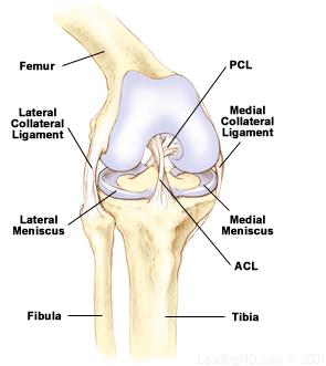
Bursa
A small, fluid-filled sac that reduces friction between adjacent structures. Examples include prepatellar and suprapatellar bursae in the knee. 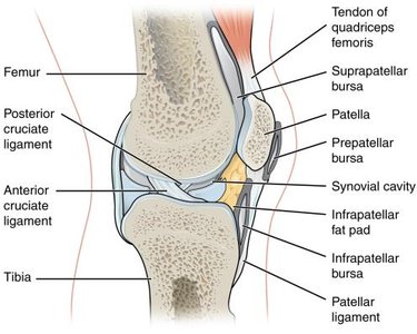
Classification of Synovial Joints by Shape
Pivot Joints
Rounded bone portion enclosed within a ring, allowing rotation (uniaxial).
Hinge Joints
Convex surface articulates with concave surface, allowing bending and straightening (uniaxial).
Condyloid Joints
Shallow depression articulates with rounded structure, allowing movement in two planes (biaxial).
Plane (Planar) Joints
Flat or slightly curved surfaces allow bones to slide across each other, with limited movement.
Saddle Joints
Saddle-shaped surfaces fit together, allowing biaxial movement (e.g., opposable thumb).
Ball-and-Socket Joints
Rounded head of one bone fits into cup-shaped depression of another, permitting the greatest range of motion (triaxial).
Movements Permitted by Synovial Joints
Flexion, Extension, and Hyperextension
Flexion: Decreases the angle between two bones.
Extension: Increases the angle between two bones.
Hyperextension: Extension beyond anatomical position.
Abduction and Adduction
Abduction: Movement away from the midline.
Adduction: Movement towards the midline.
Circumduction
Movement of a body region in a circular manner.
Rotation
Medial (internal) rotation: Anterior surface moves towards the midline.
Lateral (external) rotation: Anterior surface moves away from the midline.
Supination and Pronation
Movements of the forearm.
Supination: Palm faces anteriorly; radius and ulna are parallel.
Pronation: Palm faces posteriorly; radius and ulna are crossed.
Dorsiflexion and Plantar Flexion
Movements of the ankle joint.
Dorsiflexion: Anterior foot moves superiorly; heel remains on ground.
Plantar flexion: Posterior foot moves superiorly; toes remain on ground.
Protraction and Retraction
Anterior-posterior movements, often referring to scapula and mandible.
Elevation and Depression
Superior-inferior movements, often referring to scapula and mandible.
Summary Table: Joint Classification
Functional Classification | Structural Classification | Example |
|---|---|---|
Synarthrosis | Fibrous (Suture) | Sutures of the skull |
Synarthrosis | Fibrous (Gomphosis) | Teeth bony sockets |
Synarthrosis | Cartilaginous (Synchondrosis) | First pair of vertebrosternal ribs and sternum |
Synarthrosis | Bony fusion (Synostosis) | Epiphyseal lines of mature long bones |
Amphiarthrosis | Fibrous (Syndesmosis) | Interosseous membranes of limbs |
Amphiarthrosis | Cartilaginous (Symphysis) | Pubic symphysis, intervertebral discs |
Diarthrosis | Synovial | Shoulder, hip joints |
Clinical Relevance
Joint Movement in Physical Therapy
Physical therapists often assess and manipulate joint movement to restore function and reduce pain. Understanding joint types and permitted movements is essential for clinical practice. 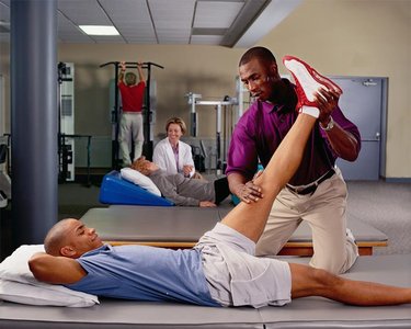
Key Terms and Definitions
Articulation: The site where two bones meet.
Synarthrosis: Immobile joint.
Amphiarthrosis: Slightly movable joint.
Diarthrosis: Freely movable joint.
Synovial fluid: Lubricating fluid in synovial joints.
Meniscus: Pad of fibrocartilage in some synovial joints.
Bursa: Fluid-filled sac reducing friction.
Equations and Formulas
Joint Range of Motion (ROM)
Range of motion is measured in degrees and depends on joint type and structure.
Biomechanical Principle
Torque at a joint: Where is torque, is force, and is the lever arm distance.
Conclusion
Joints are essential for movement and stability in the human body. Their classification, structure, and function are fundamental concepts in anatomy and physiology, with direct clinical applications in fields such as physical therapy and orthopedics. Additional info: Academic context was added to clarify joint types, movement terminology, and clinical relevance.
